FIGURE SUMMARY
Title

Skeletal muscle regeneration after extensive cryoinjury of caudal myomeres in adult zebrafish

Authors
Oudhoff, H., Hisler, V., Baumgartner, F., Rees, L., Grepper, D., Jaźwińska, A.
Source
Full text @ NPJ Regen Med

Myomere organization in adult zebrafish, and the effects of cryoinjury of the caudal peduncle.

a Left side: Schematic of the musculature displays metameric organization of somite-derived units, called myomeres. Each myomere consists of superficial muscle (red area), which comprises slow-twitch fibers, and profound muscle (brown area), which contain fast- and intermediate-twitch fibers, grouped together as fast muscles. The upper- and lower-most margins comprise longitudinal muscles, called supracarinal muscles, along the dorsal side (green) and infracarinal along the ventral midline (purple). Right side: Transversal section through the caudal peduncle shown as a schematic and F-actin staining (white). The horizontal septum separates the dorsal and ventral parts. The vertical septum separates the left and right sides of the body. Scale bar indicates 500 μm. b Coronal section through the caudal peduncle shown as a schematic and F-actin staining (white). Scale bar indicates 500 μm. c Schematic of the cryoinjury procedure shown from the ventral side of the fish. The cryoprobe was precooled in liquid nitrogen (LN2) and immediately placed on one side of the anesthetized fish for 6 s. At a specific time point after cryoinjury, fish were euthanized, and their caudal peduncles were collected for fixation. d AFOG histological staining of coronal sections at different days post-cryoinjury (dpci). The uninjured side is detected by beige staining of the muscle, whereas the cryoinjured tissue lacks this staining. Collagen is stained in blue. At 4 and 7 dpci, three to four myomeres of one side appear grayish/bluish, corresponding to the wound area. At the subsequent points, new myofibers emerge at the injured side. Scale bar indicates 500 μm. N = 4.

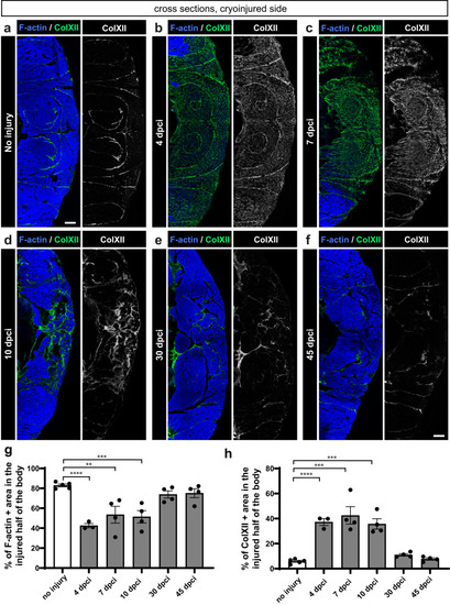
Dynamics of F-actin and Collagen XII in the wounded side of the fish body.

af One half of cross sections fluorescently stained for ColXII and F-actin. In uninjured myomeres, ColXII demarcates the myosepta, which have a complex pattern in profound muscle due to the folded structure of the myomere99. At 4, 7 and 10 dpci, ColXII accumulates within the damaged area that is devoid of F-actin. At 30 and 45 dpci, new F-actin positive muscle replaced ColXII, which persisted in the myosepta, as in the original tissue. g, h Quantification of F-actin and ColXII was performed within the entire half of the body section (cryoinjured flank). N = 3 to 5 (one fish = one dot on the graph). One-way ANOVA with Tukey’s multiple comparisons test; error bar, SEM: (ns) not significant, (*) <0.05, (**) <0.01, (***) <0.001, (****) <0.0001. Skin and dermal scales emanating fluorescence outside the muscle area were erased from images using Adobe Photoshop for clarity in this and the subsequent figures. Scale bar indicates 100 μm.

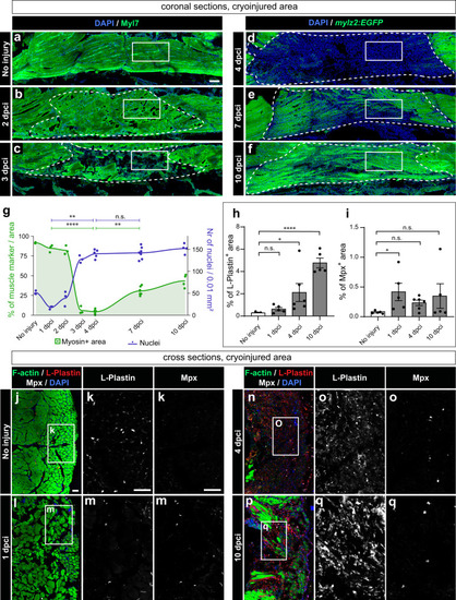
Muscle clearance is associated with cell infiltration and an immune response.

af Coronal sections fluorescently stained for four markers, of which the figure displays muscle staining (green) and nuclei (DAPI, blue), whereas two other markers (Pax7 and PCNA) are shown in (Supplementary Fig. 2 and Fig. 4). In panels (a, b, c), wild-type fish were used, and the muscle marker was MYL7 antibody, whereas in panels (d, e, f), mylz2:EGFP fish were used, and the profound muscle was detected with the GFP antibody. White frames demarcate the areas that are shown in (Fig. 4), displaying PCNA, Pax7 and DAPI. Cryoinjured areas are encircled with a dashed line, drawn based on the distorted staining of the muscle marker. For uninjured fish, a similar area was considered. g Quantification of muscle marker within the encircled cryoinjured area (left y-axis: muscle+ area, green) and cell number (right y-axis: DAPI+ nuclei, dark blue). N = 3 to 7 fish per time-point (one fish = one dot, an average of several sections per fish). Dunn’s test: (ns) not significant, (*) <0.05, (**) <0.01, (***) <0.001, (****) < 0.0001. The muscle underwent resorption at 3 and 4 dpci, which correlated with a significant increase in the number of cells within the wound area. Scale bar indicates 100 μm. Quantification of immune cell markers, L-Plastin (h) and Mpx (i) in cross sections that representatively shown below. The marker area is calculated as percentage of the cryoinjured side of the body. The measurements were performed within the entire half of the body section (cryoinjured side). N = 5 or 6 fish per time-point. Error bar = SEM. One-way ANOVA with Tukey’s multiple comparisons test. The meaning of the stars is the same as in (g). jq Representative images of uninjured control or cryoinjured side, immunostained for L-plastin and Mpx, counterstained with phalloidin (F-actin) and DAPI. The frame indicates the area that is magnified in the images to the right. Scale bar indicates 50 μm.

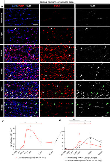
An increase of PAX7+ satellite cells and cell proliferation in the wound after muscle degeneration.

a Coronal sections fluorescently stained for four markers, of which the figure displays Pax7 (gray), PCNA (red) and DAPI (blue). The non-displayed channel is muscle staining shown in (Fig. 3a−f). The images are enlargements of the framed areas shown in (Fig. 3a−f and Supplementary Fig. 2, in which the encircled areas demarcate the region used for quantification). White arrows depict proliferating muscle stem cells (PCNA + PAX7 + DAPI+ nuclei); green arrows indicate non-proliferating muscle stem cells (PCNA negative PAX7 + DAPI+ nuclei). Scale bar indicates 50 μm. b, c Quantification of Pax7 and PCNA nuclear staining per 0.01 mm2 area of the cryoinjured area or its respective control in uninjured fish (encircled area in Supplementary Fig. 2). N = 3 to 7 fish per time-point (one fish = one dot, an average of several sections per fish). Dunn’s test: (ns) not significant, (*) <0.05, (**) <0.01, (***) <0.001, (****) <0.0001. b Dynamics of all proliferating cells (PCNA + DAPI+ nuclei, red). c Dynamics of proliferating muscle stem cells and non-proliferating muscle stem cells, as specified above.

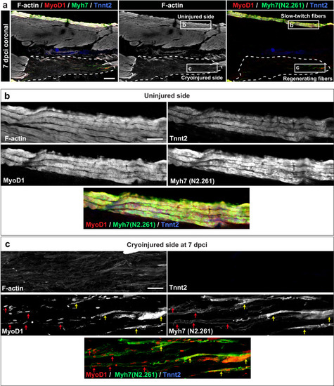
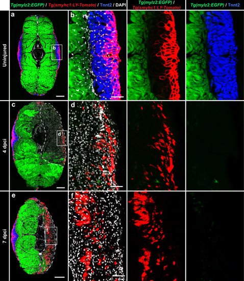
Expression of MyoD1 and slow muscle markers in early regenerating myofibers.
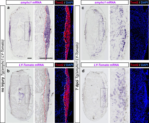
a Quadruple fluorescence staining of a coronal section at 7 dpci. A dashed line encircles the wound. The frames depict the areas that are enlarged in panels below. N = 3 fish. Scale bar indicates 200 μm. b A higher magnification of the uninjured side in the framed area as indicated in (a). The superficial fibers co-express F-actin, Tnnt2 (CT3 antibody), MyoD1 an Myh7 (N2.261 antibody). Scale bar indicates 50 μm. c A higher magnification of the cryoinjured side in the framed area as indicated in (a). The wounded area contains scattered myogenic precursors that are demarcated by nuclear MyoD1 immunoreactivity (red arrows), associated with fine Myh7 fibrils, which seem to outline an elongated morphology of cells. Near the wound margin (right side of the images), myogenic cells display cytoplasmic MyoD1 immunoreactivity and thicker Myh7 fibrils, both of which are partially overlapping (yellow arrows). Little F-actin and no Tnnt2 was detected in the wound, indicating the absence of sarcomeric tissue. Scale bar indicates 50 μm.

Comparison between smyhc1 and smyhc1:LY-Tomato expression in uninjured and regenerating muscles.

Transversal sections of smyhc1:LY-Tomato transgenic fish, stained by in-situ hybridization (purple), Tnnt2 antibody (red) and DAPI (blue). N = 3 fish per group. The frames depict the areas that are enlarged in the images to the right. Scale bar indicates 200 μm. a, b In uninjured fish, Tnnt2 positive muscle is outlined with a dashed line. The smyhc1 probe (a) labels the entire Tnnt2 positive area, corresponding to the slow muscle. The LY-Tomato probe (b) detects only the superficial-most layer of the slow muscle. c, d At 7 dpci, the smyhc1 probe (c) displays a scattered labeling in the cryoinjured side, but no Tnnt2 staining. A similar expression is detected for the LY-Tomato probe (d). Scale bar indicates 200 μm.

The smyhc1:LY-Tomato transgenic reporter is upregulated in regenerating myofibrils.

af Cross sections of the mylz2:EGFP and smyhc1:LY-Tomato double transgenic fish demarcating fast-twitch myofibers of the profound muscle (endogenous EGFP, cytosolic localization, green) and superficial layers (anti-Tomato/Cherry immunostaining, plasma membrane localization, red) of the slow muscle compartment, which is immunolabeled with Tnnt2 antibody (blue). All nuclei are stained with DAPI (white). N = 3 (control) to 5 fish (test groups), several sections per fish were analyzed. Scale bar in (a, c and e) indicates 200 μm. Scale bar in (b, d and f) indicates 50 μm. a, b In uninjured samples, a complementarity is observed between the fast muscle marker (mylz2:EGFP) and a slow muscle marker, Tnnt2. LY-Tomato immunoreactivity outlines myofibers in the outer layers of the Tnnt2-positive compartment. c, d At 4 dpci, the cryoinjured area is devoid of fast muscle (mylz2:EGFP) and slow muscle (Tnnt2) markers. In the outer layers, LY-Tomato immunoreactivity is scattered in the wound. e, f At 7 dpci, LY-Tomato immunoreactivity appears in the inner region of the damaged flank, suggesting the activation of smyhc1:LY-Tomato expression in new regenerating myofibers.

A nearly perfect restoration of fast and slow myofibers is accomplished at 30 to 45 dpci.

Higher magnifications of cross sections displaying the injured side of mylz2:EGFP and smyhc1:LY-Tomato double transgenic fish. Sections were immunostained for Tnnt2 (slow muscles) and for Tomato. Scale bar indicates 100 μm. a At 10 dpci, all three markers are activated in the wounded area, suggesting advancing regeneration. Most mylz2:EGFP positive fibers (fast myofibers) are negative for LY-Tomato and Tnnt2 (green arrows). The plasma membrane of superficial muscles is immunoreactive for the Tomato antibody (smyhc1:LY-Tomato; red arrows). Some of these cells also express Tnnt2. A few LY-Tomato positive cells are also positive for mylz2:EGFP (yellow arrows). b At 30 dpci, the wounded tissue is filled with muscle fibers. A few mylz2:EGFP single positive myofibers spread into the superficial muscle (green arrows). Some mylz2:EGFP positive myofibers also express smyhc1:LY-Tomato (yellow arrows). Most Tnnt2-positive cells are also Tomato positive cells (purple arrows). c At 45 dpci, nearly no overlap between mylz2:EGFP and smyhc1:LY-Tomato expression suggests terminal differentiation of fast and slow muscle identities. Most of mylz2:EGFP positive fibers retract from the superficial layers, and only a very few individual cells intermingle in the slow muscle compartment (green arrow). Tnnt2 positive cells (blue arrows) form a wedge-like compartment, whereas Tnnt2/LY-Tomato accumulate at the superficial layers, thus, both of which return to the original pattern. Only individual cells that are Tnnt2 or/and LY-Tomato positive remain mislocalized in the fast muscle compartment. Thus, the normal strict separation between fast and slow muscle fibers is still not perfectly re-established. dg Quantification of fluorescent areas within the wounded area (left side) compared to its respective area of the control side (right side), as indicated on the graphs. White points, uninjured side; black points, cryoinjured side. N = 3 fish, each representing an average of 3 nonadjacent sections. Error bar, SEM. Kruskal-Wallis test with Dunn’s post hoc analysis.

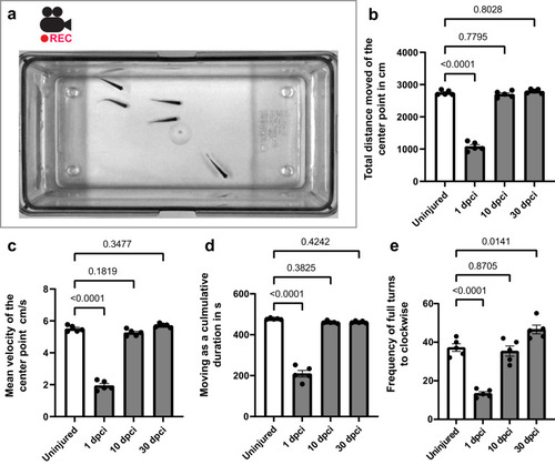
Zebrafish retain swimming activity after cryoinjury.

a A snapshot of the movie. After one minute of acclimatization in a new tank, a group of 5 fish was filmed for ten minutes. be Statistical analysis of swimming parameters, as indicated on graphs at 1, 10 and 30 dpci compared to controls using ten-minute videos. Error bar, SEM. One-way ANOVA with Dunnett’s post hoc analysis.

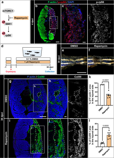
TOR signaling is required for muscle regeneration.

a Schematic illustration of mammalian TORC1 signaling with the indication of its inhibitor rapamycin. Phosphorylation of ribosomal protein S6 (p-rpS6) provides a readout for the pathway activity. b, c At 4 dpci, cross sections display p-rpS6 immunoreactivity in the wounded area. N = 3. Scale bar in (b) indicates 200 μm. Scale bar in (c) indicates 50 μm. d Experimental design with 1 µM rapamycin or 0.1% DMSO, which is the control condition. e, f Photos of the caudal peduncle. The persisting wound is encircled with a dashed line in (f). Scale bar in (e) indicates 2 mm. gj At 30 dpci, cross sections were stained for F-actin and ColXII. DMSO-treated control fish demonstrated advanced regeneration, whereas rapamycin-treated fish displayed extensive fibrotic tissue in the wound. Scale bar in (g) indicates 200 μm. Scale bar in (h) indicates 50 μm. Quantification of F-actin (k) and ColXII (l) at 30 dpci. Quantification of F-actin and ColXII was performed within the entire half of the body section (the cryoinjured lateral side). DMSO group: N = 4; rapamycin group: N = 7. Error bar, SEM. Unpaired two-tailed Student’s t test.

Acknowledgments
This image is the copyrighted work of the attributed author or publisher, and ZFIN has permission only to display this image to its users. Additional permissions should be obtained from the applicable author or publisher of the image. Full text @ NPJ Regen Med