|
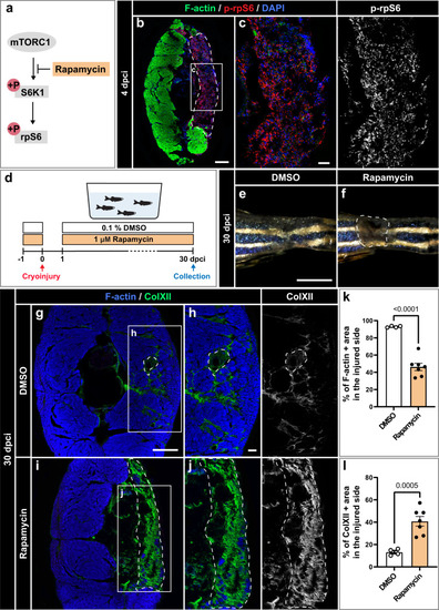
TOR signaling is required for muscle regeneration. a Schematic illustration of mammalian TORC1 signaling with the indication of its inhibitor rapamycin. Phosphorylation of ribosomal protein S6 (p-rpS6) provides a readout for the pathway activity. b, c At 4 dpci, cross sections display p-rpS6 immunoreactivity in the wounded area. N = 3. Scale bar in (b) indicates 200 μm. Scale bar in (c) indicates 50 μm. d Experimental design with 1 µM rapamycin or 0.1% DMSO, which is the control condition. e, f Photos of the caudal peduncle. The persisting wound is encircled with a dashed line in (f). Scale bar in (e) indicates 2 mm. g−j At 30 dpci, cross sections were stained for F-actin and ColXII. DMSO-treated control fish demonstrated advanced regeneration, whereas rapamycin-treated fish displayed extensive fibrotic tissue in the wound. Scale bar in (g) indicates 200 μm. Scale bar in (h) indicates 50 μm. Quantification of F-actin (k) and ColXII (l) at 30 dpci. Quantification of F-actin and ColXII was performed within the entire half of the body section (the cryoinjured lateral side). DMSO group: N = 4; rapamycin group: N = 7. Error bar, SEM. Unpaired two-tailed Student’s t test.
|