|
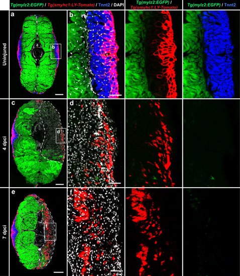
The smyhc1:LY-Tomato transgenic reporter is upregulated in regenerating myofibrils. a−f Cross sections of the mylz2:EGFP and smyhc1:LY-Tomato double transgenic fish demarcating fast-twitch myofibers of the profound muscle (endogenous EGFP, cytosolic localization, green) and superficial layers (anti-Tomato/Cherry immunostaining, plasma membrane localization, red) of the slow muscle compartment, which is immunolabeled with Tnnt2 antibody (blue). All nuclei are stained with DAPI (white). N = 3 (control) to 5 fish (test groups), several sections per fish were analyzed. Scale bar in (a, c and e) indicates 200 μm. Scale bar in (b, d and f) indicates 50 μm. a, b In uninjured samples, a complementarity is observed between the fast muscle marker (mylz2:EGFP) and a slow muscle marker, Tnnt2. LY-Tomato immunoreactivity outlines myofibers in the outer layers of the Tnnt2-positive compartment. c, d At 4 dpci, the cryoinjured area is devoid of fast muscle (mylz2:EGFP) and slow muscle (Tnnt2) markers. In the outer layers, LY-Tomato immunoreactivity is scattered in the wound. e, f At 7 dpci, LY-Tomato immunoreactivity appears in the inner region of the damaged flank, suggesting the activation of smyhc1:LY-Tomato expression in new regenerating myofibers.
|