|
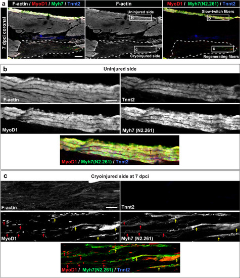
Expression of MyoD1 and slow muscle markers in early regenerating myofibers. a Quadruple fluorescence staining of a coronal section at 7 dpci. A dashed line encircles the wound. The frames depict the areas that are enlarged in panels below. N = 3 fish. Scale bar indicates 200 μm. b A higher magnification of the uninjured side in the framed area as indicated in (a). The superficial fibers co-express F-actin, Tnnt2 (CT3 antibody), MyoD1 an Myh7 (N2.261 antibody). Scale bar indicates 50 μm. c A higher magnification of the cryoinjured side in the framed area as indicated in (a). The wounded area contains scattered myogenic precursors that are demarcated by nuclear MyoD1 immunoreactivity (red arrows), associated with fine Myh7 fibrils, which seem to outline an elongated morphology of cells. Near the wound margin (right side of the images), myogenic cells display cytoplasmic MyoD1 immunoreactivity and thicker Myh7 fibrils, both of which are partially overlapping (yellow arrows). Little F-actin and no Tnnt2 was detected in the wound, indicating the absence of sarcomeric tissue. Scale bar indicates 50 μm.
|