|
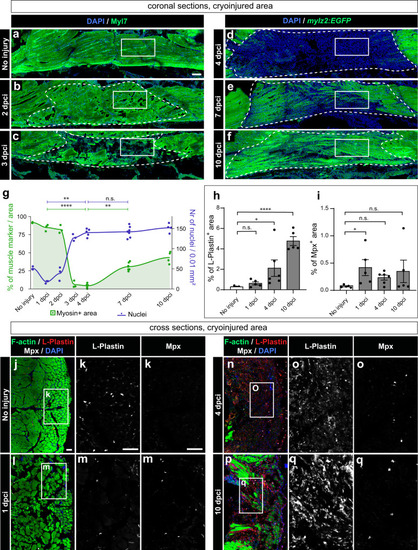
Muscle clearance is associated with cell infiltration and an immune response. a−f Coronal sections fluorescently stained for four markers, of which the figure displays muscle staining (green) and nuclei (DAPI, blue), whereas two other markers (Pax7 and PCNA) are shown in (Supplementary Fig. 2 and Fig. 4). In panels (a, b, c), wild-type fish were used, and the muscle marker was MYL7 antibody, whereas in panels (d, e, f), mylz2:EGFP fish were used, and the profound muscle was detected with the GFP antibody. White frames demarcate the areas that are shown in (Fig. 4), displaying PCNA, Pax7 and DAPI. Cryoinjured areas are encircled with a dashed line, drawn based on the distorted staining of the muscle marker. For uninjured fish, a similar area was considered. g Quantification of muscle marker within the encircled cryoinjured area (left y-axis: muscle+ area, green) and cell number (right y-axis: DAPI+ nuclei, dark blue). N = 3 to 7 fish per time-point (one fish = one dot, an average of several sections per fish). Dunn’s test: (ns) not significant, (*) <0.05, (**) <0.01, (***) <0.001, (****) < 0.0001. The muscle underwent resorption at 3 and 4 dpci, which correlated with a significant increase in the number of cells within the wound area. Scale bar indicates 100 μm. Quantification of immune cell markers, L-Plastin (h) and Mpx (i) in cross sections that representatively shown below. The marker area is calculated as percentage of the cryoinjured side of the body. The measurements were performed within the entire half of the body section (cryoinjured side). N = 5 or 6 fish per time-point. Error bar = SEM. One-way ANOVA with Tukey’s multiple comparisons test. The meaning of the stars is the same as in (g). j−q Representative images of uninjured control or cryoinjured side, immunostained for L-plastin and Mpx, counterstained with phalloidin (F-actin) and DAPI. The frame indicates the area that is magnified in the images to the right. Scale bar indicates 50 μm.
|