|
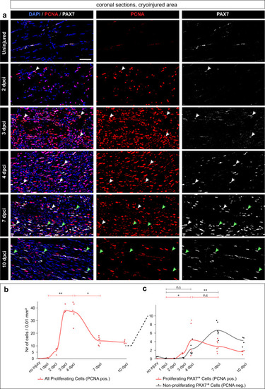
An increase of PAX7+ satellite cells and cell proliferation in the wound after muscle degeneration. a Coronal sections fluorescently stained for four markers, of which the figure displays Pax7 (gray), PCNA (red) and DAPI (blue). The non-displayed channel is muscle staining shown in (Fig. 3a−f). The images are enlargements of the framed areas shown in (Fig. 3a−f and Supplementary Fig. 2, in which the encircled areas demarcate the region used for quantification). White arrows depict proliferating muscle stem cells (PCNA + PAX7 + DAPI+ nuclei); green arrows indicate non-proliferating muscle stem cells (PCNA negative PAX7 + DAPI+ nuclei). Scale bar indicates 50 μm. b, c Quantification of Pax7 and PCNA nuclear staining per 0.01 mm2 area of the cryoinjured area or its respective control in uninjured fish (encircled area in Supplementary Fig. 2). N = 3 to 7 fish per time-point (one fish = one dot, an average of several sections per fish). Dunn’s test: (ns) not significant, (*) <0.05, (**) <0.01, (***) <0.001, (****) <0.0001. b Dynamics of all proliferating cells (PCNA + DAPI+ nuclei, red). c Dynamics of proliferating muscle stem cells and non-proliferating muscle stem cells, as specified above.
|