|
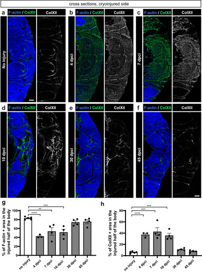
Dynamics of F-actin and Collagen XII in the wounded side of the fish body. a−f One half of cross sections fluorescently stained for ColXII and F-actin. In uninjured myomeres, ColXII demarcates the myosepta, which have a complex pattern in profound muscle due to the folded structure of the myomere99. At 4, 7 and 10 dpci, ColXII accumulates within the damaged area that is devoid of F-actin. At 30 and 45 dpci, new F-actin positive muscle replaced ColXII, which persisted in the myosepta, as in the original tissue. g, h Quantification of F-actin and ColXII was performed within the entire half of the body section (cryoinjured flank). N = 3 to 5 (one fish = one dot on the graph). One-way ANOVA with Tukey’s multiple comparisons test; error bar, SEM: (ns) not significant, (*) <0.05, (**) <0.01, (***) <0.001, (****) <0.0001. Skin and dermal scales emanating fluorescence outside the muscle area were erased from images using Adobe Photoshop for clarity in this and the subsequent figures. Scale bar indicates 100 μm.
|