- Title
-
Annexin-A5 and annexin-A6 silencing prevents metastasis of breast cancer cells in zebrafish
- Authors
- Gounou, C., Bouvet, F., Liet, B., Prouzet-Mauléon, V., d'Agata, L., Harté, E., Argoul, F., Siegfried, G., Iggo, R., Khatib, A.M., Bouter, A.
- Source
- Full text @ Biol. Cell
|
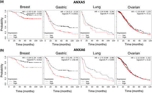
High ANXA5 or ANXA6 expression in advanced cancer patients is a poor prognosis factor. (a,b) Survival curves from Kaplan–Meier plot profiles for breast (n = 201), gastric (n = 305), lung (n = 70), or ovarian (n = 1220) high-grade and -stage cancer patients, stratified by high (red line) and low expression (black line) of ANXA5 (a) or ANXA6 (b) mRNA (Affymetrix identifier, 200782_at and 200982_s_at, respectively). In each analysis, hazard ratio (HR) and log-rank probability were calculated. A HR significantly different from 1 means that survival was better in one of both groups. For example, if a HR is 1.5, the survival probability in group 1 is 1.5 higher than in group 2. To note, cancer grade (typically numbered from 1 to 3) correlates with the velocity of the disease development while stage (from 1 to 4) characterizes the extent of affected tissues. Advanced cancers were triple-negative subtype, stage 3, stage 3 + grade 4, and stage 3, for breast, lung, ovarian, and gastric cancer, respectively. HR, hazard ratio. |
|
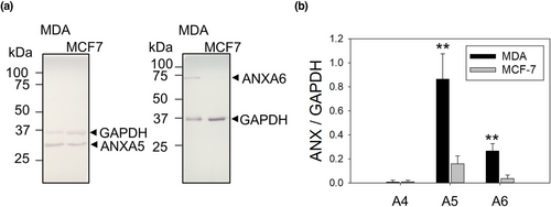
Invasiveness properties in breast cancer cells are correlated with high expression of ANXA5 and ANXA6. 10 μg of protein extracts from MDA-MB-231 and MCF7 cells were analyzed by western-blotting. (a) Representative image of western-blot analysis showing the revelation of ANXA5 and ANXA6 in MDA-MB-231 and MCF7 cells, compared to GAPDH (loading control). The whole membranes are presented in Figure S2. (b) The histogram presents mean values (± SEM) of the ratio ANX/GAPDH from five independent experiments, analyzed by the gel analysis plugging of ImageJ. Student t-test for independent samples. **p < 0.01. |
|
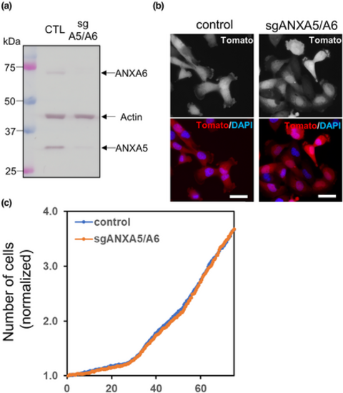
sgANXA5/A6 MDA-MB-231 cells exhibit morphology and growth properties similar to control cells. (a) ANXA5/A6 deficient MDA-MB-231 cells were established by the CRIPSR-Cas9 strategy. The cellular content of ANXA5 and ANXA6 in sgANXA5/A6 MDA-MB-231 cells was quantified by western-blotting. The expression of ANXA5 and ANXA6 is decreased of more than 95% and 85% in sgANXA5/A6 MDA-MB-231 cells, respectively. (b) Control and sgANXA5/A6 MDA-MB-231 cells, which expressed constitutively the tdTomato fluorescent protein (red), were imaged by fluorescence microscopy. Scale bars = 40 μm. (c) Proliferation of control or sgANXA5/A6 MDA-MB-231 cells was monitored with a lensfree microscope enabling long-term observation of a large cell population (Allier et al., 2017). The averaged number of cells (+/− SEM) normalized to the number of seeded cells from three independent experiments is presented. |
|
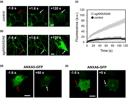
ANXA5 and ANXA6 are involved in membrane repair of MDA-MB-231 cells. (a,b) Sequences of representative images showing the response of a control (a) or sgANXA5/A6 (b) MDA-MB-231 cell to a membrane damage performed by 110-mW infrared laser irradiation, in the presence of FM1-43 (green). In all figures, the area of membrane irradiation is marked with a red arrow before irradiation and a white arrow after irradiation. Scale bars: 10 μm. (c) Kinetic data represent the FM1−43 fluorescence intensity integrated over whole cell sections, averaged for about 30 cells (+/− SEM). For a majority of control MDA-MB-231 cells, the fluorescence intensity reached a plateau after about 60 s (black filled circles). For sgANXA5/A6 MDA-MB-231 cells, a continuous and large increase of the fluorescence intensity was observed (empty circles), indicating the absence of membrane resealing. (d,e) Recruitment of ANXA5-GFP (d) and ANXA6-GFP (e) to the site of membrane injury. Red arrow, area before irradiation; white arrow, area after irradiation. After laser injury, MDA-MB-231 cells exhibited an accumulation of ANXA5 and ANXA6 at the disruption site. Scale bars: 20 μm. |
|
Silencing of ANXA5 and ANXA6 prevents metastasis process of MDA-MB-231 cells in zebrafish. (a,b) Control (a, n = 24) or sgANXA5/A6 MDA-MB-231 (b, n = 24) cells were injected in the perivitelline space of Casper zebrafish embryos. Tumor imaging was done by fluorescence microscopy at 24- and 48-hpi through the tdTomato fluorescent protein constitutively expressed in MDA-MB-231 cells and merged with bright-field images. Insets display magnified images of a portion of the tail and head with metastases (white arrows). (c) The percentage of embryos, either injected by control or sgANXA5/A6 MDA-MB-231 cells, which presented caudal or head metastases was quantified at 24 hpi. Mortality rate was calculated at 48 hpi. |


