
Fig. 4
- ID
- ZDB-FIG-231106-35
- Publication
- Gounou et al., 2023 - Annexin-A5 and annexin-A6 silencing prevents metastasis of breast cancer cells in zebrafish
- Other Figures
- All Figure Page
- Back to All Figure Page
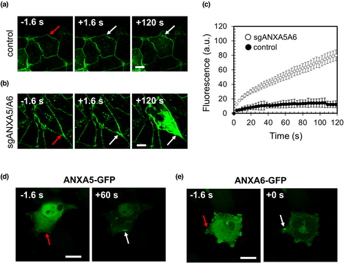
ANXA5 and ANXA6 are involved in membrane repair of MDA-MB-231 cells. (a,b) Sequences of representative images showing the response of a control (a) or sgANXA5/A6 (b) MDA-MB-231 cell to a membrane damage performed by 110-mW infrared laser irradiation, in the presence of FM1-43 (green). In all figures, the area of membrane irradiation is marked with a red arrow before irradiation and a white arrow after irradiation. Scale bars: 10 μm. (c) Kinetic data represent the FM1−43 fluorescence intensity integrated over whole cell sections, averaged for about 30 cells (+/− SEM). For a majority of control MDA-MB-231 cells, the fluorescence intensity reached a plateau after about 60 s (black filled circles). For sgANXA5/A6 MDA-MB-231 cells, a continuous and large increase of the fluorescence intensity was observed (empty circles), indicating the absence of membrane resealing. (d,e) Recruitment of ANXA5-GFP (d) and ANXA6-GFP (e) to the site of membrane injury. Red arrow, area before irradiation; white arrow, area after irradiation. After laser injury, MDA-MB-231 cells exhibited an accumulation of ANXA5 and ANXA6 at the disruption site. Scale bars: 20 μm. |