
|
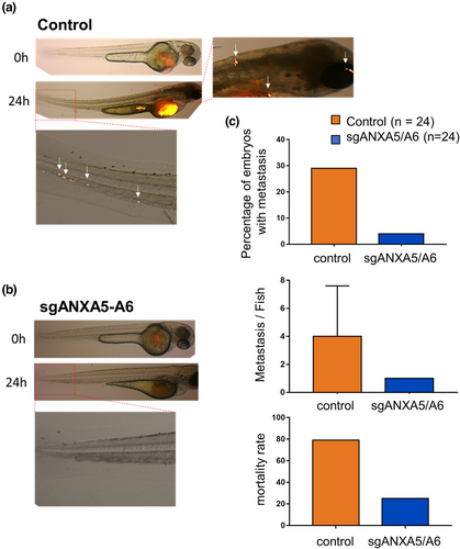
Fig. 5 Silencing of ANXA5 and ANXA6 prevents metastasis process of MDA-MB-231 cells in zebrafish. (a,b) Control (a, n = 24) or sgANXA5/A6 MDA-MB-231 (b, n = 24) cells were injected in the perivitelline space of Casper zebrafish embryos. Tumor imaging was done by fluorescence microscopy at 24- and 48-hpi through the tdTomato fluorescent protein constitutively expressed in MDA-MB-231 cells and merged with bright-field images. Insets display magnified images of a portion of the tail and head with metastases (white arrows). (c) The percentage of embryos, either injected by control or sgANXA5/A6 MDA-MB-231 cells, which presented caudal or head metastases was quantified at 24 hpi. Mortality rate was calculated at 48 hpi.