
- Title
-
Ipconazole Disrupts Mitochondrial Homeostasis and Alters GABAergic Neuronal Development in Zebrafish
- Authors
- Lee, G., Banik, A., Eum, J., Hwang, B.J., Kwon, S.H., Kee, Y.
- Source
- Full text @ Int. J. Mol. Sci.
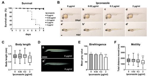
Ipconazole alters larval locomotive behavior in zebrafish development. (A) Survival rate (%) of zebrafish larvae treated with ipconazole, repeated six times (n = 300 each); (B) lateral views of the bright field imaging of the embryos at 25 h post fertilization (hpf) (a–d) and 4 days post fertilization (dpf) (a’–d’), repeated four times (n = 40 each), scale bar, 500 μm; (C) body length of the larvae treated with ipconazole at 4 dpf; (D) lateral views of 3-dpf larvae subjected to polarized light microscopy (a,a’), scale bar, 500 μm; (E) quantitation of the average birefringence of the larval muscle, repeated three times (n = 30 each); (F) locomotor activity of 6-dpf larvae treated with ipconazole. Graphical representation of the average distance traveled in the dark condition. * p < 0.05, ** p < 0.01, *** p < 0.001. |
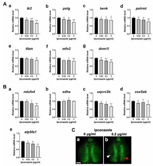
Ipconazole disrupts mitochondrial genome maintenance and respiratory complexes. (A) mRNA profiling of the genes involved in the mitochondrial DNA replication and transcription, tk2 (a), polg (b), twnk (c), polrmt (d), and mfn2) (f), and a fission marker (dnm1l) (g); (B) mRNA profiling of mitochondrial respiratory complex genes: ndufs4 of Complex I (a), sdha of Complex II (b), uqcrc2b of Complex III (c), cox5ab of Complex IV (d), and atp5fa1 of Complex V (e); (C) confocal images of mitochondria (in green) in the midbrain of 3-dpf Tg(Xla.Eef1a1:mlsEGFP)cms1 larvae treated with ipconazole at 0 μg/mL (a), and 0.2 μg/mL (b). Scale bar, 50 μm; white arrowhead, neuropil; red arrowhead, periventricular zone. * p < 0.05, ** p < 0.01, *** p < 0.001. |
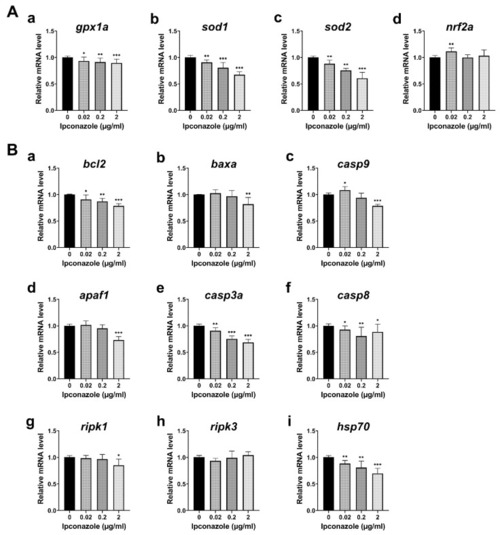
Ipconazole reduces expressions of antioxidant enzymes, caspases, and hsp70. (A) RT-PCR (real-time PCR) shows the antioxidant genes were reduced in the 28-hpf embryos treated with ipconazole at 0, 0.02, 0.2, and 2 μg/mL: gpx1a (a), sod1 (b) sod2 (c), and nrf2 (d); (B) mRNA expression of the apoptotic genes, bcl2 (a), baxa (b), casp9 (c), apaf1 (d), casp3a (e), and casp8 (f), and the necroptotic genes, ripk1 (g) and ripk3 (h), as well as the molecular chaperon hsp70 (i) were monitored. * p < 0.05, ** p < 0.01, *** p < 0.001. |
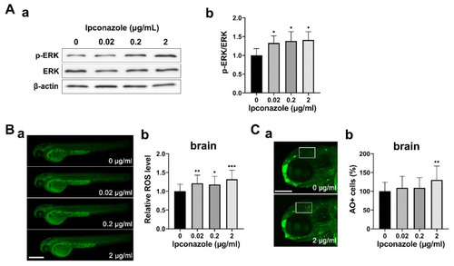
Ipconazole activates ERK1/2 signaling under oxidative stress. (A) Phosphorylation of ERK1/2 was increased in 28-hpf embryos treated with ipconazole. (a) Western blots using the antibodies against p-ERK, ERK, and β-actin proteins, respectively. (b) Graph of the quantitative analysis; (B) reactive oxygen species (ROS) were generated in the 48-hpf embryos treated with ipconazole. Scale bar, 500 µm. (a) Lateral views of the embryos subjected to DCFH-DA (2′,7′-dichlorodihydrofluorescein diacetate) staining and fluorescence imaging. (b) ROS level was quantitated by measuring the fluorescence intensity of ROS in the brain using the ImageJ software; (C) fluorescently labeled apoptotic cells in the brain (white box) of the 72-hpf ipconazole-treated embryos were counted using acridine orange staining. (a) Lateral views of the head subjected to acridine orange staining. Scale bar, 200 μm. (b) Graph of the quantitative analysis. * p < 0.05, ** p < 0.01, *** p < 0.001. |
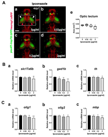
Ipconazole preferentially alters the development of GABAergic neurons. (A) Dorsal views of the brain of the 4-dpf larvae of Tg(pou4f1-hsp70l: GFP)rw0110b and Tg(-8.4neurog1:nRFP)sb3 transgenic fish using confocal microscopy (a–d) and the size analysis of the optic tectum in the embryos treated with ipconazole (e). White box, optic tectum (OT); white arrowhead, neuropil; red arrowhead, periventricular layer, scale bar, 100 μm; (B) mRNA expressions of the following neuronal markers in the 3-dpf embryos using RT-PCR: slc17a6b for excitatory glutamatergic neurons (a), gad1b for GABAergic inhibitory neurons (b), and th for dopaminergic neurons (c); (C) mRNA expressions of the following glial cell markers: olig1 and olig2 for oligodendrocyte formation (a,b) and mbp for myelination (c) in the 3-dpf embryos. * p < 0.05, ** p < 0.01, *** p < 0.001. |
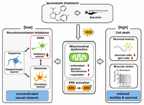
Potential mechanism of neurodevelopmental toxicity of ipconazole pesticide in zebrafish. The zebrafish embryos treated with ipconazole generate ROS and show evidence of dose-dependent mitochondrial dysfunction and ERK1/2 activation. Ipconazole specifically inhibited the development of GABAergic inhibitory neurons at low doses. In contrast, glutamatergic and dopaminergic neurons remained unaffected, possibly disrupting the interplay among the major neurotransmission systems and uncoordinated neural functions in the central nervous system. High concentrations of ipconazole cause cell death in neuronal and muscle development, reducing the motility and survival of embryos. *** p < 0.001. |