
|
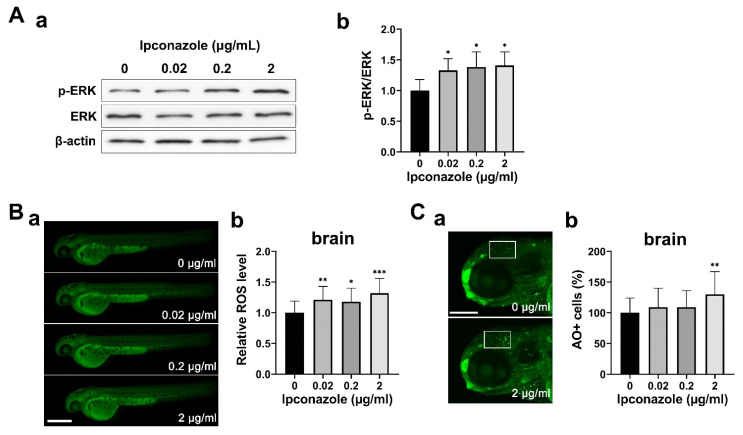
Figure 4 Ipconazole activates ERK1/2 signaling under oxidative stress. (A) Phosphorylation of ERK1/2 was increased in 28-hpf embryos treated with ipconazole. (a) Western blots using the antibodies against p-ERK, ERK, and β-actin proteins, respectively. (b) Graph of the quantitative analysis; (B) reactive oxygen species (ROS) were generated in the 48-hpf embryos treated with ipconazole. Scale bar, 500 µm. (a) Lateral views of the embryos subjected to DCFH-DA (2′,7′-dichlorodihydrofluorescein diacetate) staining and fluorescence imaging. (b) ROS level was quantitated by measuring the fluorescence intensity of ROS in the brain using the ImageJ software; (C) fluorescently labeled apoptotic cells in the brain (white box) of the 72-hpf ipconazole-treated embryos were counted using acridine orange staining. (a) Lateral views of the head subjected to acridine orange staining. Scale bar, 200 μm. (b) Graph of the quantitative analysis. * p < 0.05, ** p < 0.01, *** p < 0.001.