- Title
-
Early calcium and cardiac contraction defects in a model of phospholamban R9C mutation in zebrafish
- Authors
- Vicente, M., Salgado-Almario, J., Valiente-Gabioud, A.A., Collins, M.M., Vincent, P., Domingo, B., Llopis, J.
- Source
- Full text @ J. Mol. Cell. Cardiol.
|
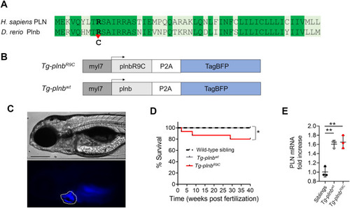
Generation of Tg-plnbwt and Tg-plnbR9C zebrafish lines. A) Alignment of the human PLN and zebrafish Plnb protein sequences. Identical amino acids are indicated in dark green. The bold type indicates the Arg9Cys mutation point. B) myl7:plnbR9C-P2A-TagBFP and myl7:plnb-P2A-TagBFP transgenes flanked by Tol2 sequences (not shown in the scheme). C) Transmitted and fluorescence images of a representative 4 dpf Tg-plnbR9C zebrafish larva showing the expression of TagBFP in the heart. The scale bar represents 200 μm. D) Kaplan-Meier curve showing the survival of wild-type siblings, Tg-plnbwt and Tg-plnbR9C fish up to 40 weeks. A Log-rank (Mantel-Cox) test was used. E) Quantitative PCR plot showing the relative expression levels of plnb in 3 dpf Tg-plnbwt and Tg-plnbR9C larvae, compared to wild-type siblings (n = 3 pools of ten hearts each). GADPH was used for normalization. Statistical analysis was performed using a one-way ANOVA test with Tukey's multiple comparisons post-test. (* p < 0.05, ** p < 0.01). (For interpretation of the references to colour in this figure legend, the reader is referred to the web version of this article.) |
|
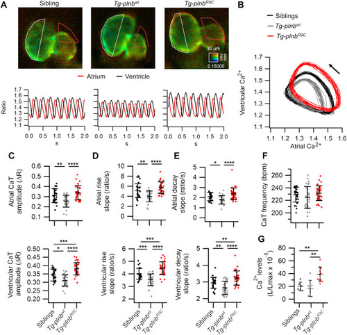
Atrial and ventricular Ca2+ handling in sibling, Tg-plnbwt and Tg-plnbR9C 3 dpf zebrafish larvae expressing mCyRFP1-GCaMP6f (A-F) or GFP-Aequorin (G). A) Ratiometric images from beating hearts and their corresponding atrial and ventricular Ca2+ traces (ratio) from representative sibling, Tg-plnbwt and Tg-plnbR9C larvae. B) Diagram of the ventricular vs. atrial Ca2+ levels from the larvae in A. The arrow indicates the direction of time; ∼18 cardiac cycles are shown. C) Atrial and ventricular Ca2+ transient amplitude (CaT), D) atrial and ventricular Ca2+ rise slopes, E) atrial and ventricular Ca2+ decay slopes, F) frequency of the CaT (HR) from sibling (n = 34), Tg-plnbwt (n = 31) and Tg-plnbR9C (n = 34) zebrafish larvae. G) Time-averaged ventricular Ca2+ levels (L/Lmax) from sibling (n = 6), Tg-plnbwt (n = 7) and Tg-plnbR9C (n = 11) zebrafish larvae expressing GFP-Aequorin reconstituted with diacetyl h-coelenterazine; images were acquired at 1 frame/s. Statistical analysis was performed using a one-way ANOVA test with Tukey's multiple comparisons post-test. Data are shown as mean ± SD (* p < 0.05, ** p < 0.01, *** p < 0.001, **** p < 0.0001). The microscopy chamber temperature was 28 °C. |
|
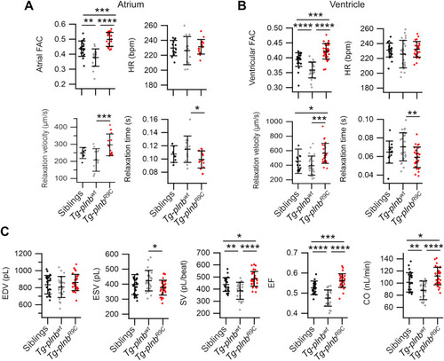
Atrial and ventricular contractility and hemodynamics in sibling, Tg-plnbwt and Tg-plnbR9C 3 dpf zebrafish larvae expressing mCyRFP1-GCaMP6f. Fractional area change (FAC) for the atrium (A) and the ventricle (B) from sibling (n = 23 for A and 30 for B), Tg-plnbwt (n = 18 for A and 24 for B) and Tg-plnbR9C (n = 19 for A and 32 for B) zebrafish larvae. Heart rate (HR), relaxation velocity and relaxation time 90%–10% for the atrium (A) and the ventricle (B) from sibling (n = 13 for A and 30 for B), Tg-plnbwt (n = 12 for A and 24 for B) and Tg-plnbR9C (n = 15 for A and 32 for B) zebrafish larvae. C) Ventricular function parameters: EDV, ESV, SV, EF, and CO in sibling (n = 30), Tg-plnbwt (n = 24) and Tg-plnbR9C (n = 32) zebrafish larvae. Statistical analysis was performed using a one-way ANOVA test with Tukey's multiple comparisons post-test. Data are shown as mean ± SD, (* p < 0.05, ** p < 0.01, *** p < 0.001, **** p < 0.0001). |
|
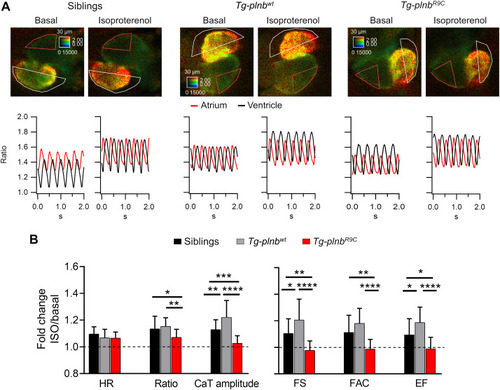
Tg-plnbR9C larvae displayed a blunted β-adrenergic response at 3 dpf. A) Ratiometric images from beating hearts (taken at ventricular systole) and their corresponding atrial and ventricular Ca2+ traces from representative sibling, Tg-plnbwt and Tg-plnbR9C larvae in basal condition and after 30-min incubation in 100 μM isoproterenol. B) Fold change of isoproterenol over basal values of the HR, average ratio (Ca2+ level), CaT amplitude, FS, FAC and EF in the ventricle of sibling (n = 24), Tg-plnbwt (n = 17) and Tg-plnbR9C (n = 19) larvae. Statistical analysis was performed using a one-way ANOVA test with Tukey's multiple comparisons post-test. Data are shown as mean ± SD (* p < 0.05, ** p < 0.01, *** p < 0.001, **** p < 0.0001). The microscopy chamber temperature was set at 25 °C. |
|
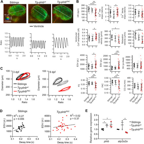
Cardiac Ca2+ handling and contractility in sibling, Tg-plnbwt and Tg-plnbR9C 9 dpf zebrafish larvae expressing mCyRFP1-GCaMP6f. A) Ratiometric images from beating hearts and their corresponding ventricular Ca2+ traces from representative sibling, Tg-plnbwt and Tg-plnbR9C larvae. B) HR, ventricular CaT amplitude, ventricular Ca2+ rise and decay rates, contraction and relaxation velocities, ESV, EDV, SV, FS, FAC and CO in sibling (n = 26), Tg-plnbwt (n = 17) and Tg-plnbR9C zebrafish larvae (n = 31). Statistical analysis was performed using a one-way ANOVA test with Tukey's multiple comparisons post-test. C) Diagrams of the ventricular diameter (μm) vs. ventricular Ca2+ levels (ratio) from the 3 and 9 dpf representative sibling, Tg-plnbwt and Tg-plnbR9C larvae shown in A. D) Pearson's correlation test between the decay time of CaT and EDV from 9 dpf sibling (n = 26) and Tg-plnbR9C (n = 31) zebrafish larvae. E) Relative expression levels (qPCR) of plnb and atp2a2a in 9 dpf Tg-plnbwt, Tg-plnbR9C and sibling larvae (n = 3, 3 and 4 pools of ten hearts each, respectively). GADPH was used as a normalization control. Data are shown as mean ± SD, (* p < 0.05, ** p < 0.01, *** p < 0.001, **** p < 0.0001). |
|
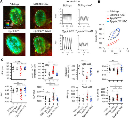
Cardiac Ca2+ handling and contractility in NAC-treated sibling and Tg-plnbR9C 9 dpf zebrafish larvae expressing mCyRFP1-GCaMP6f. A) Ratiometric images from beating hearts, with their corresponding ventricular Ca2+ traces from representative control and NAC-treated sibling and Tg-plnbR9C larvae. B) Diagram of the ventricular diameter (μm) vs. ventricular Ca2+ levels (ratio) from representative control and NAC-treated sibling and Tg-plnbR9C larvae. C) HR, ventricular CaT amplitude, relaxation velocity, FS, FAC, ESV, EDV and SV from control and NAC-treated sibling (n = 23 and n = 21, respectively), and control and NAC-treated Tg-plnbR9C (n = 29 and n = 27, respectively) zebrafish larvae. Statistical analysis was performed using a one-way ANOVA test with Tukey's multiple comparisons post-test. Data are shown as mean ± SD, (* p < 0.05, ** p < 0.01, *** p < 0.001, **** p < 0.0001). |
|
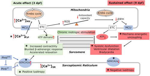
Summary of the pathological model for R9C mutation. PLNR9C mutation leads to increased Ca2+ levels, contractility, and a blunted β-adrenergic response in the heart, consistent with the observations in 3 dpf larvae. The chronic inotropic stimulation caused by R9C, and the energetic demand exerts a pathological effect displaying ventricular dilation, systolic dysfunction, bradycardia and negative lusitropy, as observed in 9 dpf larvae. A reduced antioxidant capacity may alter the redox balance in line with the observation in 9 dpf treated with NAC . Asterisks indicate the findigs shown in this work; the mitochondrial changes are a working model (ETC, electron transport chain; MCU, mitochondrial Ca2+ uniporter; ROS, reactive oxygen species). |
Reprinted from Journal of Molecular and Cellular Cardiology, 173, Vicente, M., Salgado-Almario, J., Valiente-Gabioud, A.A., Collins, M.M., Vincent, P., Domingo, B., Llopis, J., Early calcium and cardiac contraction defects in a model of phospholamban R9C mutation in zebrafish, 127-140, Copyright (2022) with permission from Elsevier. Full text @ J. Mol. Cell. Cardiol.

