Fig. 2
- ID
- ZDB-FIG-230505-6
- Publication
- Vicente et al., 2022 - Early calcium and cardiac contraction defects in a model of phospholamban R9C mutation in zebrafish
- Other Figures
- All Figure Page
- Back to All Figure Page
|
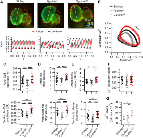
Atrial and ventricular Ca2+ handling in sibling, Tg-plnbwt and Tg-plnbR9C 3 dpf zebrafish larvae expressing mCyRFP1-GCaMP6f (A-F) or GFP-Aequorin (G). A) Ratiometric images from beating hearts and their corresponding atrial and ventricular Ca2+ traces (ratio) from representative sibling, Tg-plnbwt and Tg-plnbR9C larvae. B) Diagram of the ventricular vs. atrial Ca2+ levels from the larvae in A. The arrow indicates the direction of time; ∼18 cardiac cycles are shown. C) Atrial and ventricular Ca2+ transient amplitude (CaT), D) atrial and ventricular Ca2+ rise slopes, E) atrial and ventricular Ca2+ decay slopes, F) frequency of the CaT (HR) from sibling (n = 34), Tg-plnbwt (n = 31) and Tg-plnbR9C (n = 34) zebrafish larvae. G) Time-averaged ventricular Ca2+ levels (L/Lmax) from sibling (n = 6), Tg-plnbwt (n = 7) and Tg-plnbR9C (n = 11) zebrafish larvae expressing GFP-Aequorin reconstituted with diacetyl h-coelenterazine; images were acquired at 1 frame/s. Statistical analysis was performed using a one-way ANOVA test with Tukey's multiple comparisons post-test. Data are shown as mean ± SD (* p < 0.05, ** p < 0.01, *** p < 0.001, **** p < 0.0001). The microscopy chamber temperature was 28 °C. |
Reprinted from Journal of Molecular and Cellular Cardiology, 173, Vicente, M., Salgado-Almario, J., Valiente-Gabioud, A.A., Collins, M.M., Vincent, P., Domingo, B., Llopis, J., Early calcium and cardiac contraction defects in a model of phospholamban R9C mutation in zebrafish, 127-140, Copyright (2022) with permission from Elsevier. Full text @ J. Mol. Cell. Cardiol.

