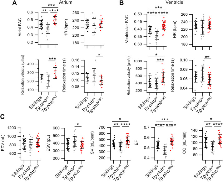
Fig. 3 Atrial and ventricular contractility and hemodynamics in sibling, Tg-plnbwt and Tg-plnbR9C 3 dpf zebrafish larvae expressing mCyRFP1-GCaMP6f. Fractional area change (FAC) for the atrium (A) and the ventricle (B) from sibling (n = 23 for A and 30 for B), Tg-plnbwt (n = 18 for A and 24 for B) and Tg-plnbR9C (n = 19 for A and 32 for B) zebrafish larvae. Heart rate (HR), relaxation velocity and relaxation time 90%–10% for the atrium (A) and the ventricle (B) from sibling (n = 13 for A and 30 for B), Tg-plnbwt (n = 12 for A and 24 for B) and Tg-plnbR9C (n = 15 for A and 32 for B) zebrafish larvae. C) Ventricular function parameters: EDV, ESV, SV, EF, and CO in sibling (n = 30), Tg-plnbwt (n = 24) and Tg-plnbR9C (n = 32) zebrafish larvae. Statistical analysis was performed using a one-way ANOVA test with Tukey's multiple comparisons post-test. Data are shown as mean ± SD, (* p < 0.05, ** p < 0.01, *** p < 0.001, **** p < 0.0001).
Reprinted from Journal of Molecular and Cellular Cardiology, 173, Vicente, M., Salgado-Almario, J., Valiente-Gabioud, A.A., Collins, M.M., Vincent, P., Domingo, B., Llopis, J., Early calcium and cardiac contraction defects in a model of phospholamban R9C mutation in zebrafish, 127-140, Copyright (2022) with permission from Elsevier. Full text @ J. Mol. Cell. Cardiol.

