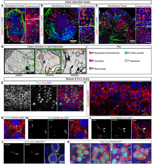
Fig. 6. The zygotene cilium is conserved in zebrafish male meiosis and mouse oogenesis. (A to C) Seminiferous tubules of adult zebrafish testes labeled with AcTub and Sycp3 [(A), n = 35 tubules in n = 3 testes] or Telo-FISH [(B), n = 29 tubules in n = 2 testes] or γH2Ax [(C), n = 34 tubules in n = 3 testes]. Left panels: optical sections of entire tubules (white outline; scale bars, 20 μm). Right panels: magnified images of zygotene bouquet spermatocytes (early zygotene bouquet in top panels, and late zygotene bouquet in bottom panels; scale bars, 10 μm). L/Z, leptotene/zygotene primary spermatocytes; P, pachytene spermatocytes; E1, initial spermatids; E2, intermediate spermatids; E3, final (mature) spermatids. (D) TEM images of adult testis showing ciliary structures (see arrowheads in key) within (left panels) prophase spermatocytes (synapsed chromosomes, pink arrowheads). Colored panels are magnifications of color-coded boxes. Magnifications and scale bars are as indicated. n = 25 spermatocytes in n = 2 testes. (E) Mouse E14.5 ovaries labeled with Vasa and Sycp3. Scale bar, 20 μm. (F) Mouse E14.5 ovaries labeled with Vasa and AcTub showing AcTub-positive CBs (pink arrows) and cilia (white arrows) in Vasa-labeled cysts. n = 4 ovaries. Scale bar, 15 μm. (G to J) E14.5 ovaries labeled with AcTub, Vasa, and γTub [(G), n = 2 ovaries], or Arl13b (H), n = 2 ovaries], or GluTub [(I), n = 2 ovaries], or Adcy3 [(J), n = 2 ovaries]. Scale bars, 5 μm. (K) E14.5 ovaries co-labeled with Vasa, Sycp3, GluTub, and DAPI. Individual channel images are shown in fig. S18 (n = 2 ovaries). Scale bars, 5 μm. In (E) to (J), cilia (white arrows) and CB-like structures (pink arrows) are indicated.
|


