
- Title
-
The role of gfi1.2 in the development of zebrafish inner ear
- Authors
- Yu, R., Wang, P., Chen, X.W.
- Source
- Full text @ Hear. Res.
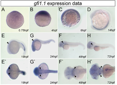
The expression of gfi1.1 during early development in zebrafish was detected by in situ hybridization. The animal pole is at the top in A–C. The anterior aspect is at the top and the dorsal at the right in D. The anterior side is at the left and the dorsal side at the top in E–H. A lateral view is shown in E–H and the dorsal view in E'–H'. Arrowheads indicate the domains of hair cells of the two maculae and three cristae in the ear. EXPRESSION / LABELING:
|

The expression of gfi1.2 during early development in zebrafish was detected by in situ hybridization. The animal pole is toward the top in A–C. The anterior is toward the top and the dorsal is facing right in D. The anterior is toward the left and dorsal is toward the top in E–H. A lateral view is shown in E–H and the dorsal view in E'–H'. Arrowheads indicate the domains of hair cells of the two maculae and three cristae in the ear. EXPRESSION / LABELING:
|
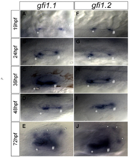
A higher magnification of gfi1.1 and gfi1.2 expression in the hair cells of maculae and cristae. A maculae consists of both utricle and saccule. Utricle is labeled by *, saccule is labeled by #, and cristae is labeled by triangle. EXPRESSION / LABELING:
|
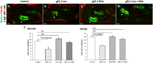
The number of hair cells and neurons was significantly decreased by MO-mediated gfi1.2 knockdown and can be rescued by exogenous gfi1.2. Neurons were labeled by the immunostaining of Myo6 and tubulin at 2 dpf. The number of neurons in the zebrafish neuromast was compared in (A) wild-type (wt), (B) MO-mediated gfi1.2-knockdown, (C) gfi1.2 mRNA-overexpressing, and (D) MO gfi1.2 + gfi1.2 mRNA. Hair cells were also labeled by the immunostaining of Myo6 and tubulin at 2 dpf. The number of hair cells in the zebrafish inner ear was compared in (E) wild-type (wt), (F) MO-mediated gfi1.2-knockdown, (G) gfi1.2 mRNA-overexpressing, and (H) MO gfi1.2 + gfi1.2 mRNA. In the gfi1.2 mutants, hair cells in the utricle maculae (UM) and saccule maculae (SM) significantly decreased in number (p < 0.05). Over-expression of gfi1.2 in the wild-type zebrafish did not affect the number of hair cell number (E). NS: not significant. The utricle is indicated by a white *, and the contralateral structure is the saccule. PHENOTYPE:
|
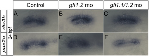
In situ hybridization assay of the otic specification and patterning-related markers was performed using pax2a and dlx3b probes (lateral view). The expression was similar in the gfi1.2 mutant and gfi1.1/gfi1.2 mutant embryos at 24 hpf. |
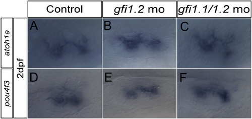
In situ hybridization assay of the hair cell differentiation and maturation-related markers was performed using pou4f3 and Atoh1a probes (lateral view). The expression was similar in the gfi1.2 mutant and gfi1.1/gfi1.2 mutant embryos at 24 hpf. |
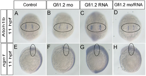
In situ hybridization assay of the pro-neural markers was performed using atoh1b probes (dorsal view) and neurog1 probes (lateral view). The expression of both markers was detected in the wild-type and mutant embryos at 11 hpf. EXPRESSION / LABELING:
PHENOTYPE:
|
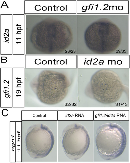
Correlations among gfi1.2, id2a, and neurog1 detected by in situ hybridization. (A) Expression of id2a in wild-type and mutant embryos at 11 hpf. (B) Expression of gfi1.2 in wild-type and id2a mutant embryos at 19 hpf. (C) Exogenous overexpression of gfi1.2 rescued the reduced expression of neurog1 caused by id2a. |
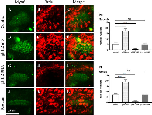
Myo6 (green) is the marker for hair cells, and BrdU positivity (red) represents proliferating cells. Merged images labeled proliferating hair cells in the specimen. The presence of proliferating hair cells in wild-type zebrafish (control group) was demonstrated in (A–C). With gfi1.2 MO treatment, the proliferating hair cells increased in number (D–F). On the other hand, gfi1.2 mRNA treatment suppressed the proliferation of hair cells (G–I). gfi1.2 mRNA-induced hair cell suppression was rescued by adding gfi1.2 MO (J–L). Proliferating hair cells in (M) saccule and (N) utricle are further quantified. (For interpretation of the references to color in this figure legend, the reader is referred to the web version of this article.) PHENOTYPE:
|
Reprinted from Hearing Research, 396, Yu, R., Wang, P., Chen, X.W., The role of gfi1.2 in the development of zebrafish inner ear, 108055, Copyright (2020) with permission from Elsevier. Full text @ Hear. Res.