
- Title
-
MGST1, a GSH transferase/peroxidase essential for development and hematopoietic stem cell differentiation
- Authors
- Bräutigam, L., Zhang, J., Dreij, K., Spahiu, L., Holmgren, A., Abe, H., Tew, K.D., Townsend, D.M., Kelner, M.J., Morgenstern, R., Johansson, K.
- Source
- Full text @ Redox Biol.
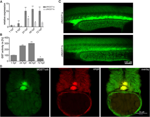
(A) Relative expression of zfMGST1a/b during zebrafish embryonic development. (B) Relative activity of GST enzymes during zebrafish embryonic development. (C) Expression of MGST1a and MGST1b in the intermediate cell mass of 24 hpf embryos. (D) MGST1a/b protein colocalizes with cmyb, a marker specific for HSCs, in 48 hpf embryos. Confocal images were taken on a Leica LSM700 with a 20× lens; Alexa 488 and 555 filters were used; images stacks were produced with ImageJ, GIMP was used to adjust the gamma channel. EXPRESSION / LABELING:
|
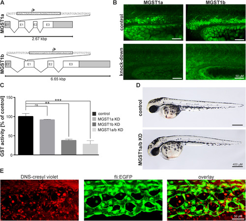
Morpholino induced knock-down of MGST1 in zebrafish. (A) Genomic organization of zfMGST1 and location of morpholino attachment sites targeting transcription. (B) Immunohistochemistry in embryos injected with morpholinos knocking down MGST1a/b. (C) Enzymatic activity of GST enzymes in extracts of embryos injected with morpholinos knocking-down MGST1a, MGST1b or both. (D) Gross morphology of embryos injected with morpholinos knocking-down MGST1a/b. (E) DNS-cresyl violet staining indicating MGST1 positive cells in the caudal hematopoietic tissue, see also Supplementary movie S1. Brightfield images were taken on a Leica MZ16 microscope equipped with a Leica DFC300FX camera; Confocal images were taken on a Leica LSM700 with a 20× lens; Alexa 488 and 555 filters were used; images stacks were produced with ImageJ, GIMP was used to adjust the gamma channel. PHENOTYPE:
|
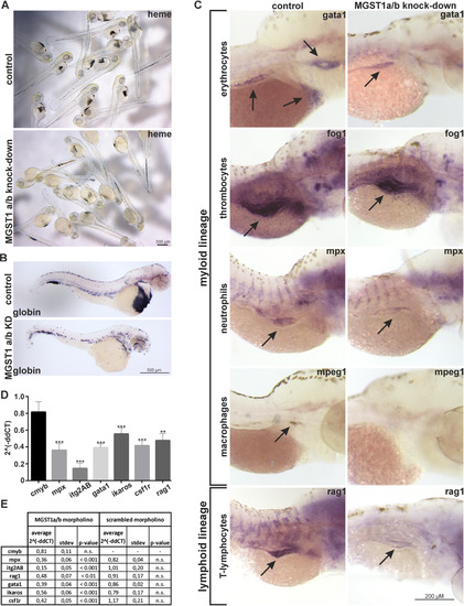
Loss of functional MGST1 blocks differentiation of hematopoietic stem cells in zebrafish. (A) Histochemical staining of hemoglobin in living zebrafish embryos. (B) Whole-mount in situ hybridization in 48 hpf embryos staining globin transcripts specific for erythrocytes. (C) Whole-mount in situ hybridization against marker genes for different myloid and lymphoid lineages in 96 hpf embryos. (D) RTqPCR quantifying transcripts specific for different myloid and lymphoid lineages in 96 hpf embryos. (E) Quantification and statistics for RTqPCR. Brightfield images were taken on a Leica MZ16 microscope equipped with a Leica DFC300FX camera; images stacks were produced with ImageJ, GIMP was used to adjust the gamma channel. |
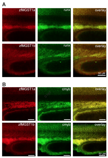
Whole-mount immunohistochemistry against MGST1a/b and cmyb / runx in 48 hpf embryos. EXPRESSION / LABELING:
|
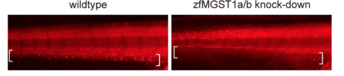
48 hour old embryos stained with the fluorogenic GST substrate 2,4-dinitrobenzene sulfonamide cresyl violet. The region of the intermediate cell mass is marked with brackets. Images were taken with excitation at 540 nm and emission at 620 nm. |
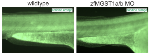
Acridine orange staining for the detection of cells undergoing cell death in 48 hpf embryos injected with MGST1a/b morpholino. |