
- Title
-
Morphofunctional Alterations in Zebrafish (Danio rerio) Gills after Exposure to Mercury Chloride
- Authors
- Macirella, R., Brunelli, E.
- Source
- Full text @ Int. J. Mol. Sci.

D. rerio gill apparatus in basal conditions: (A) SEM micrographs showing concentric microridges of pavement cells (PVCs). F = filament and L = lamellae. Bar 50 µm; (B) light micrographs in toluidine blue showing general morphological organization of branchial epithelium; F = filament and L = lamellae. Bar 50 µm. |
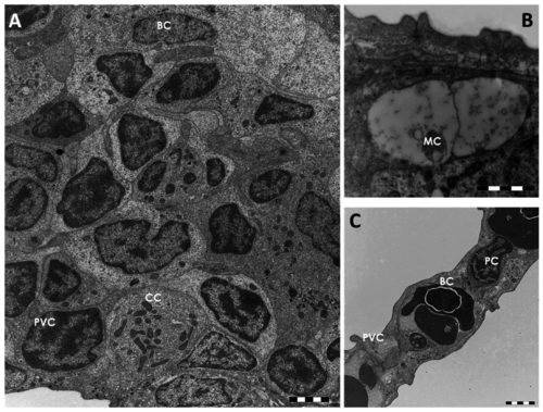
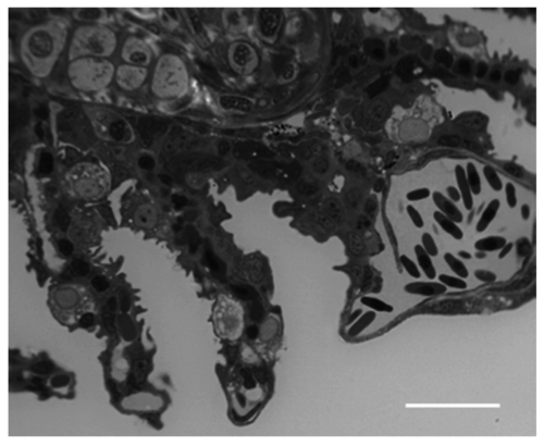
TEM micrographs of the gill apparatus in D. rerio under basal conditions: (A) cellular organization in the primary epithelium. CC = chloride cell, PVC = pavement cell; BC = basal cell; (B) high magnification of a mucous cell (MC); (C) ultrastructural organization of the secondary epithelium. PVC = pavement cell; BC = basal cell; PC = pillar cell. All bars 2 µm. |
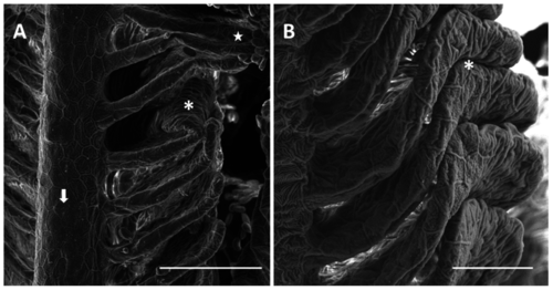
SEM micrographs of the primary and secondary epithelium in D. rerio after 96 h of exposure to 7.7 µg/L of HgCl2: (A) degeneration in PVCs microridges in the primary epithelium (arrow); folding in the distal portion of lamellae (asterisk) and lamellar fusion (star). Bar 50 µm; (B) higher magnification of lamellar fusion (asterisk). Bar 20 µm. |

Sagittal section in toluidine blue of the gill apparatus in D. rerio after 96 h of exposure to 7.7 µg/L of HgCl2: (A) detachment of epithelium from connective tissue that create wide lacunae in gills lamellae (asterisk). Bar 20 µm; (B) hypertrophy in endothelial cells and blood congestion (black arrow). Bar 50 µm. |
|
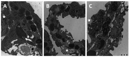
TEM micrographs of the gill apparatus in D. rerio after 96 h of exposure to 7.7 µg/L of HgCl2: (A) early degeneration in the inner layers of filaments; (B) detachment of epithelium (asterisk) and hypertrophy of chloride cell (CC); (C) high magnification of detachment in secondary epithelium (asterisk) and degeneration of PVCs (star); (D) loss of PVCs connections with the basal lamina (asterisk). All bars 2 µm. 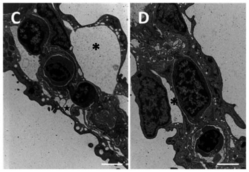
|
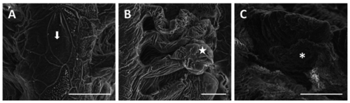
SEM micrographs of the gill apparatus in D. rerio after 96 h of exposure to 38.5 µg/L of HgCl2: (A) degeneration of microridges in primary epithelium (arrow). Bar 20 µm; (B) wrinkled surface of primary and secondary epithelium with swelling in the distal portion of lamellae (star). Bar 20 µm; (C) increase in mucous secretion (asterisk). Bar 100 µm. |
Cross section in toluidine blue of the gill apparatus in D. rerio after 96 h of exposure to 38.5 µg/L of HgCl2: hyperplasia of secondary epithelium with the appearance of CCs and MCs. Bar 20 µm. |
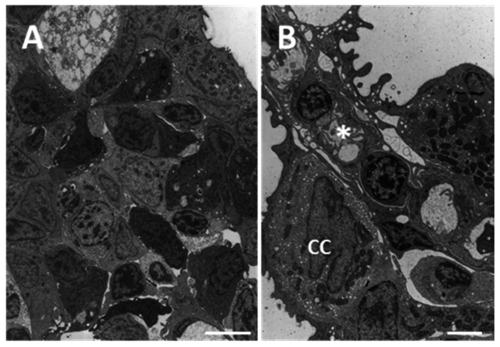
TEM micrographs of the gill apparatus in D. rerio after 96 h of exposure to 38.5 µg/L of HgCl2: (A) degeneration in primary epithelium with degeneration of mucous cells (asterisk), epithelial gaps (arrow), and macrophage infiltrations (star); (B) folding of secondary lamellae, hypertrophic mucous cell (asterisk), and tissue hyperplasia; (C) appearance of CCs in secondary lamellae and formation of deep invagination in CC (star). All bars 2 µm. |
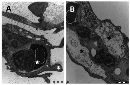
TEM micrographs of the gill apparatus in D. rerio after 96 h of exposure to 38.5 µg/L of HgCl2: (A) high magnification of long processes in PVCs (asterisks) and edema formation in the distal portion of lamellae (star); (B) degenerated and apoptotic PVCs (asterisks) with the disappearance of PCs. All bars 2 µm. |
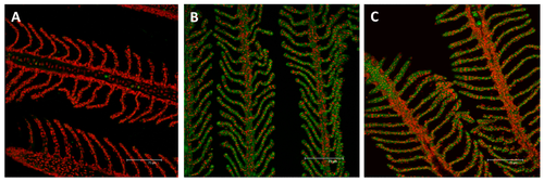
Confocal micrographs of the D. rerio gill apparatus. Sections labeled with a mouse monoclonal antibody against metallothionein (MT) (green–Fluorescein Isothiocyanate (FITC) labeled); nuclei labeled with propidium iodide (red); (A) no MTs expression in the gills of the control group; (B) after 96 h of exposure to 7.7 µg/L of HgCl2, MTs immunoreactivity strongly appear in both the primary and secondary epithelium; (C) after 96 h of exposure to 38.5 µg/L of HgCl2, the intensity of staining lightly decrease compared to the basal condition in both the filament and lamellar epithelium. All bars 75 µm. |

Confocal micrographs of D. rerio gill apparatus. Sections labeled with a mouse monoclonal antibody against Na+/K+-ATPase (green–FITC labeled); nuclei labeled with propidium iodide (red); (A) detection of Na+/K+-ATPase in the CCs of the interlamellar region in the basal condition; (B) after 96 h of exposure to 7.7 µg/L of HgCl2, Na+/K+-ATPase immunoreactivity strongly decrease compared to the basal condition but the fluorescence labeling appears at the level of the secondary epithelium; (C) After 96 h of exposure to 38.5 µg/L of HgCl2, the expression for Na+/K+-ATPase increase in both the filament and lamellar epithelium. All bars 75 µm. |