
FIGURE
Fig. 8
Fig. 8
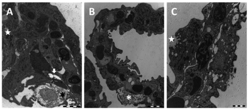
TEM micrographs of the gill apparatus in D. rerio after 96 h of exposure to 38.5 µg/L of HgCl2: (A) degeneration in primary epithelium with degeneration of mucous cells (asterisk), epithelial gaps (arrow), and macrophage infiltrations (star); (B) folding of secondary lamellae, hypertrophic mucous cell (asterisk), and tissue hyperplasia; (C) appearance of CCs in secondary lamellae and formation of deep invagination in CC (star). All bars 2 µm. |
Expression Data
Expression Detail
Antibody Labeling
Phenotype Data
Phenotype Detail
Acknowledgments
This image is the copyrighted work of the attributed author or publisher, and
ZFIN has permission only to display this image to its users.
Additional permissions should be obtained from the applicable author or publisher of the image.
Full text @ Int. J. Mol. Sci.