
Image
|
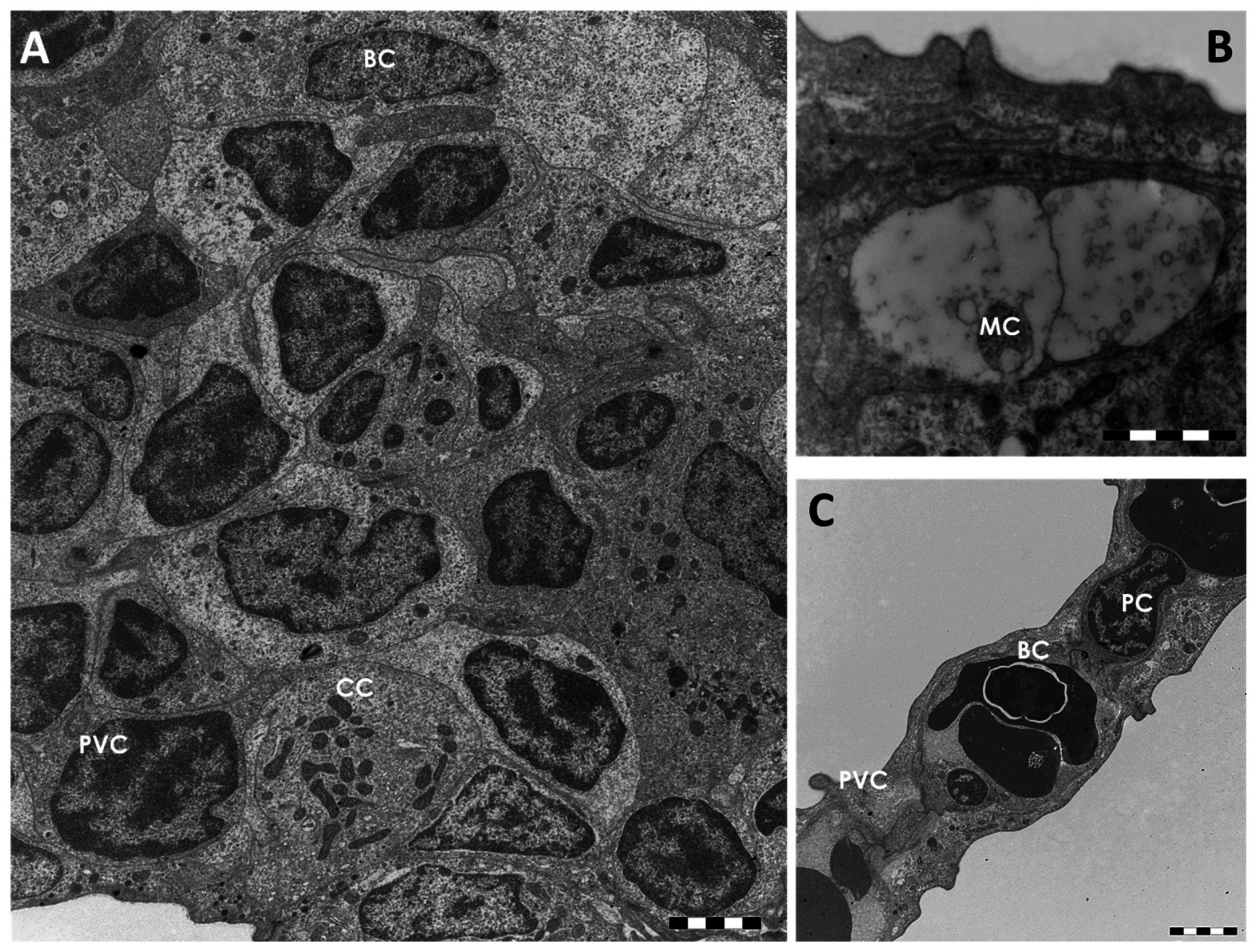
Figure Caption
Fig. 2
TEM micrographs of the gill apparatus in D. rerio under basal conditions: (A) cellular organization in the primary epithelium. CC = chloride cell, PVC = pavement cell; BC = basal cell; (B) high magnification of a mucous cell (MC); (C) ultrastructural organization of the secondary epithelium. PVC = pavement cell; BC = basal cell; PC = pillar cell. All bars 2 µm.
Acknowledgments
This image is the copyrighted work of the attributed author or publisher, and
ZFIN has permission only to display this image to its users.
Additional permissions should be obtained from the applicable author or publisher of the image.
Full text @ Int. J. Mol. Sci.