
|
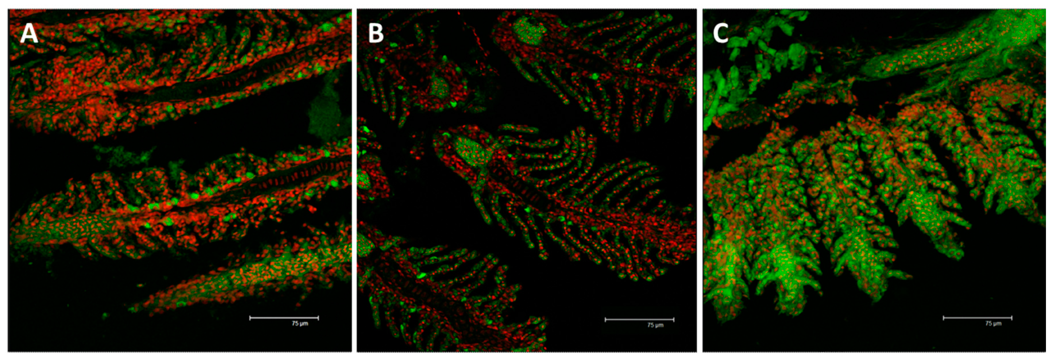
Fig. 12
Confocal micrographs of D. rerio gill apparatus. Sections labeled with a mouse monoclonal antibody against Na+/K+-ATPase (green–FITC labeled); nuclei labeled with propidium iodide (red); (A) detection of Na+/K+-ATPase in the CCs of the interlamellar region in the basal condition; (B) after 96 h of exposure to 7.7 µg/L of HgCl2, Na+/K+-ATPase immunoreactivity strongly decrease compared to the basal condition but the fluorescence labeling appears at the level of the secondary epithelium; (C) After 96 h of exposure to 38.5 µg/L of HgCl2, the expression for Na+/K+-ATPase increase in both the filament and lamellar epithelium. All bars 75 µm.