
Image
|
Figure Caption
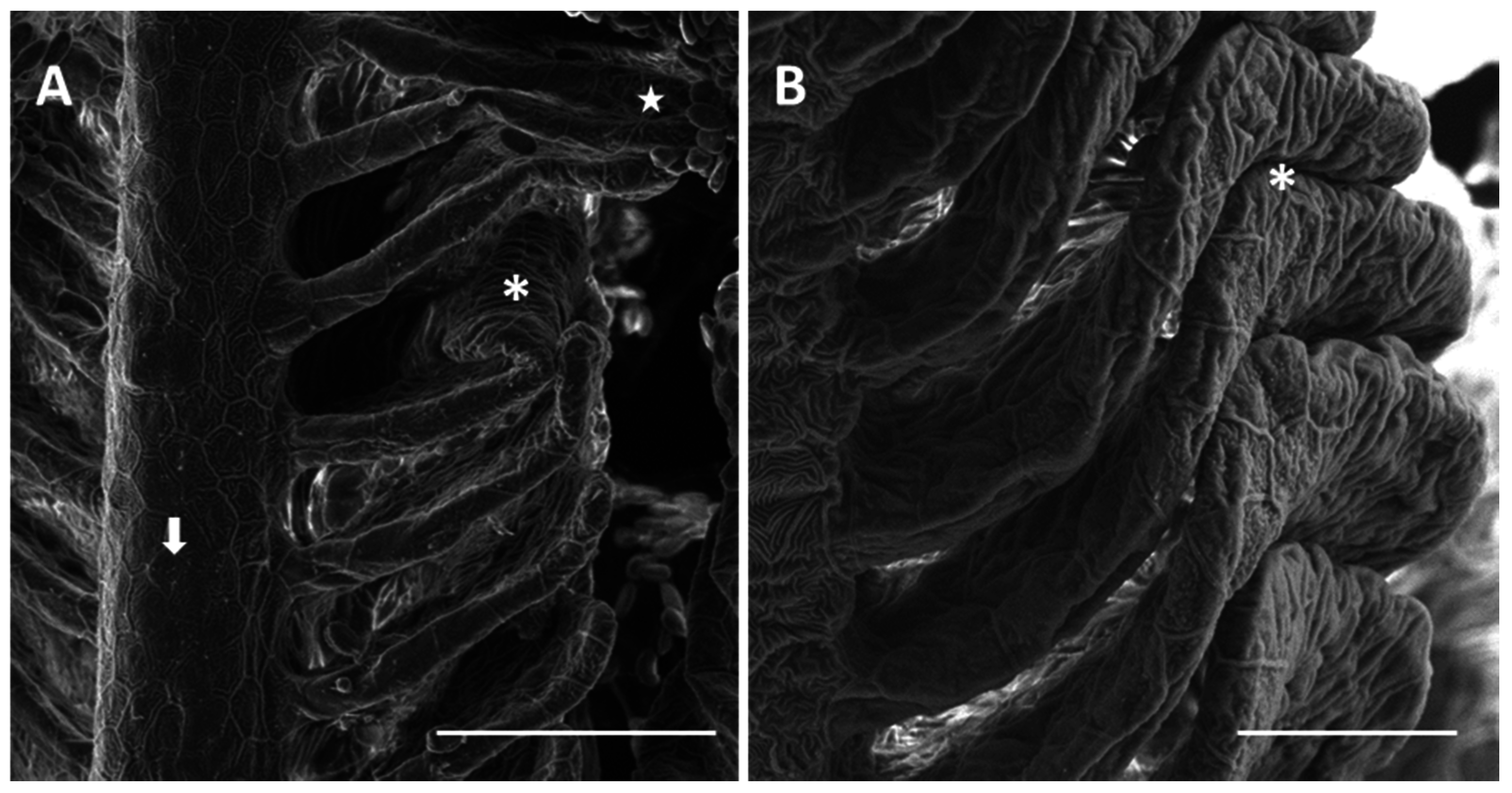
Fig. 3
SEM micrographs of the primary and secondary epithelium in D. rerio after 96 h of exposure to 7.7 µg/L of HgCl2: (A) degeneration in PVCs microridges in the primary epithelium (arrow); folding in the distal portion of lamellae (asterisk) and lamellar fusion (star). Bar 50 µm; (B) higher magnification of lamellar fusion (asterisk). Bar 20 µm.
Acknowledgments
This image is the copyrighted work of the attributed author or publisher, and
ZFIN has permission only to display this image to its users.
Additional permissions should be obtained from the applicable author or publisher of the image.
Full text @ Int. J. Mol. Sci.