
FIGURE
Fig. 6
Fig. 6
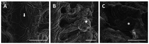
SEM micrographs of the gill apparatus in D. rerio after 96 h of exposure to 38.5 µg/L of HgCl2: (A) degeneration of microridges in primary epithelium (arrow). Bar 20 µm; (B) wrinkled surface of primary and secondary epithelium with swelling in the distal portion of lamellae (star). Bar 20 µm; (C) increase in mucous secretion (asterisk). Bar 100 µm. |
Expression Data
Expression Detail
Antibody Labeling
Phenotype Data
Phenotype Detail
Acknowledgments
This image is the copyrighted work of the attributed author or publisher, and
ZFIN has permission only to display this image to its users.
Additional permissions should be obtained from the applicable author or publisher of the image.
Full text @ Int. J. Mol. Sci.