
Image
|
Figure Caption
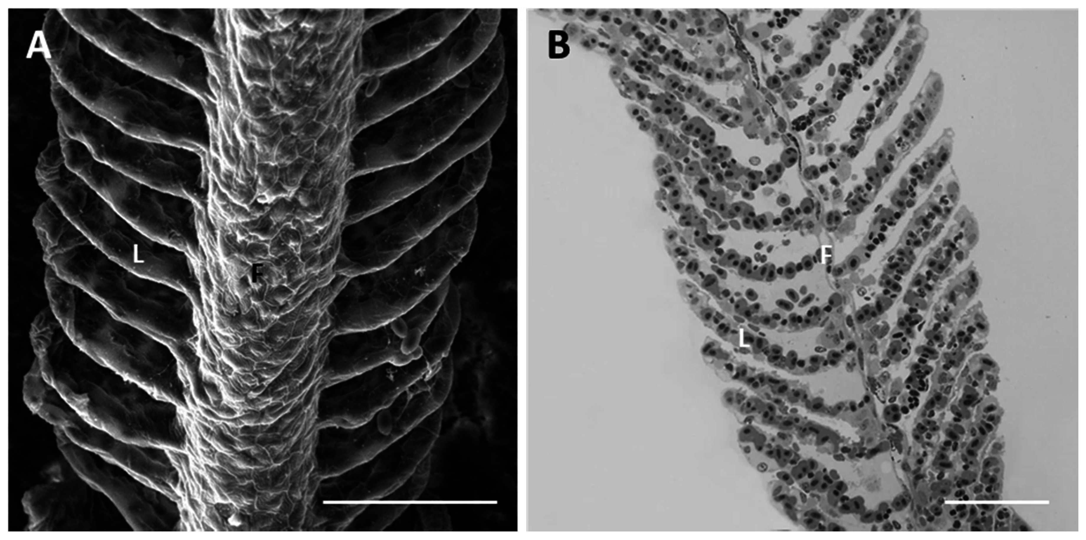
Fig. 1
D. rerio gill apparatus in basal conditions: (A) SEM micrographs showing concentric microridges of pavement cells (PVCs). F = filament and L = lamellae. Bar 50 µm; (B) light micrographs in toluidine blue showing general morphological organization of branchial epithelium; F = filament and L = lamellae. Bar 50 µm.
Acknowledgments
This image is the copyrighted work of the attributed author or publisher, and
ZFIN has permission only to display this image to its users.
Additional permissions should be obtained from the applicable author or publisher of the image.
Full text @ Int. J. Mol. Sci.