
Image
|
Figure Caption
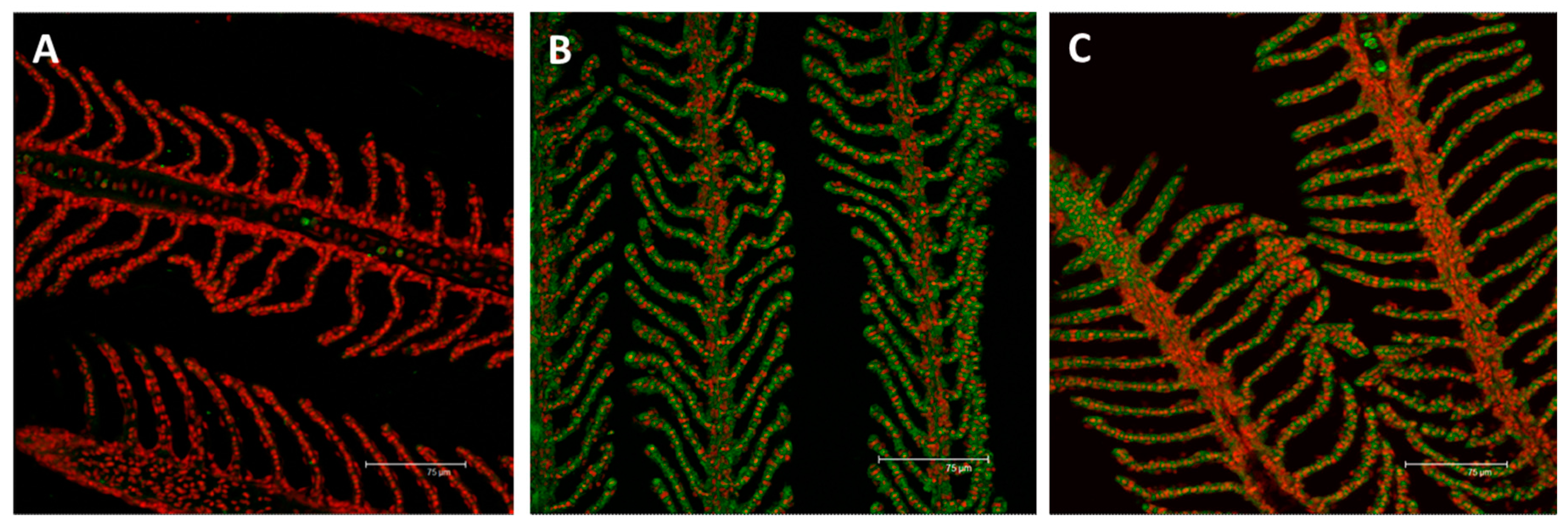
Fig. 10
Confocal micrographs of the D. rerio gill apparatus. Sections labeled with a mouse monoclonal antibody against metallothionein (MT) (green–Fluorescein Isothiocyanate (FITC) labeled); nuclei labeled with propidium iodide (red); (A) no MTs expression in the gills of the control group; (B) after 96 h of exposure to 7.7 µg/L of HgCl2, MTs immunoreactivity strongly appear in both the primary and secondary epithelium; (C) after 96 h of exposure to 38.5 µg/L of HgCl2, the intensity of staining lightly decrease compared to the basal condition in both the filament and lamellar epithelium. All bars 75 µm.
Acknowledgments
This image is the copyrighted work of the attributed author or publisher, and
ZFIN has permission only to display this image to its users.
Additional permissions should be obtained from the applicable author or publisher of the image.
Full text @ Int. J. Mol. Sci.