- Title
-
Akt1 Mediates Neuronal Differentiation in Zebrafish via a Reciprocal Interaction with Notch Signaling
- Authors
- Cheng, Y.C., Hsieh, F.Y., Chiang, M.C., Scotting, P.J., Shih, H.Y., Lin, S.J., Wu, H.L., and Lee, H.T.
- Source
- Full text @ PLoS One
|
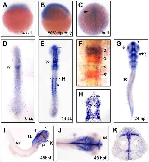
akt1 expression in the developing zebrafish. akt1 expression was detected by in situ hybridization in the developing nervous system during zebrafish embryogenesis. The embryo stages are shown in the bottom right corner of each panel. (C–F and G) Dorsal view with anterior to the top. (I) Lateral view with anterior to the right. (J) Dorsal view with anterior to the right. akt1 expression appears first in the developing nervous system during the bud stage (arrowhead in C) and later becomes restricted to specific brain areas (D–K). Relatively weak expression was detected in the spinal cord from the bud stage (C) and persisted until the final stage that was analyzed (48 hpf) (C–E and G–J). (F) akt1 expression (purple) in rhombomere 2 analyzed by double in situ hybridization with krox20 (red). H and K are cross-sections from E and I, respectively. hb, hindbrain; le, lens; mhb, midbrain-hindbrain boundary; p, pallium; pr, pharyngeal arches; r2–r5, rhombomere 2–5; s, somites; sc, spinal cord; t, tectum; tel, telencephalon. |
|
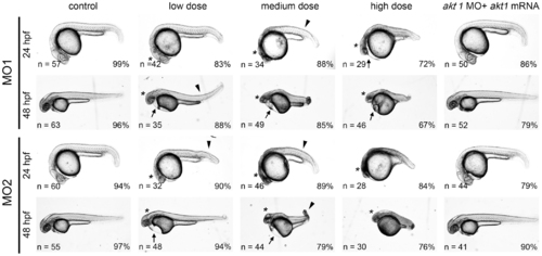
Akt1 knockdown using specific morpholinos is sufficient to cause developmental abnormalities in a dose-dependent manner. Representative images of morpholino-injected embryos. The following doses were used for microinjection: low dose, 6 ng MO1 or 1 ng MO2; medium dose, 10 ng MO1 or 2 ng MO2; high dose, 14 ng MO1 or 4 ng MO2. Arrowheads denote bent body characteristics, while arrows indicate edema in the pericardial sac. Brain malformations are marked by asterisks. These morphological phenotypes were rescued by concomitant injection with 1 ng akt1 mRNA, as shown in the right panels. |
|
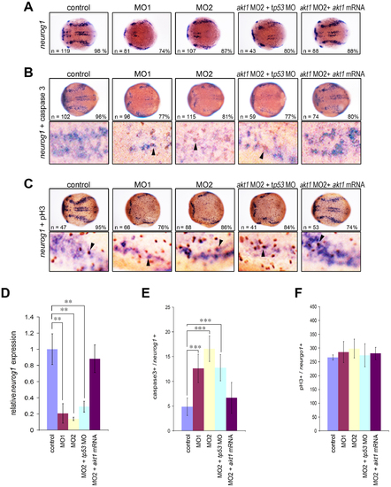
Decreased neuronal precursors in Akt1 knockdown embryos during early neurogenesis. (A) In situ hybridization showing the dramatic downregulation of neurogenin1 in akt1 morpholino-injected embryos. This effect was rescued by co-injection with akt1 mRNA but not a tp53 morpholino, which was confirmed by qPCR analysis (D). (B) Apoptotic cells were detected using caspase-3 antibody (brown) and double-labeled with neurogenin1 (purple), which showed an increase in apoptotic cells in akt1 morphants. The double-labeled cells are indicated with arrows and quantified in E. (C) Proliferating neurons were double-labeled with phosphohistone H3 antibody and neurogenin1 riboprobes (arrows), which showed no significant difference between akt1 morphants and controls. This was confirmed by quantification (F). In A–C the lower panels are enlargements of the upper panels. **, p<0.01; ***, p<0.001. |
|
Akt1 morphants exhibit upregulation of HuC/D expression with simultaneous downregulation of neurogenin1 expression. (A) During the bud stage, injection with akt1 morpholinos upregulated the expression of elavl3 compared with the controls. (B) HuC/D-expressing post-mitotic neurons increased massively in akt1 morpholino-injected embryos, as shown by immunohistochemical analysis with anti-Hu antibody at 24 hpf. Note the ectopic HuC/D expression in the ventricular zone (arrows in insert panels). (C) At the same stage, neurogenin1 expression was reduced significantly by Akt1 knockdown. (D) Levels of HuC/D expression were confirmed by Western blot analysis and are quantified in the top panel. (E) qPCR analysis further confirmed the decreased expression of neurogenein1 at 24 hpf (C) in akt1 morpholino-injected embryos. All phenotypes could be rescued by concomitant injection with akt1 mRNA but not a tp53 morpholino. **, p<0.01. |
|
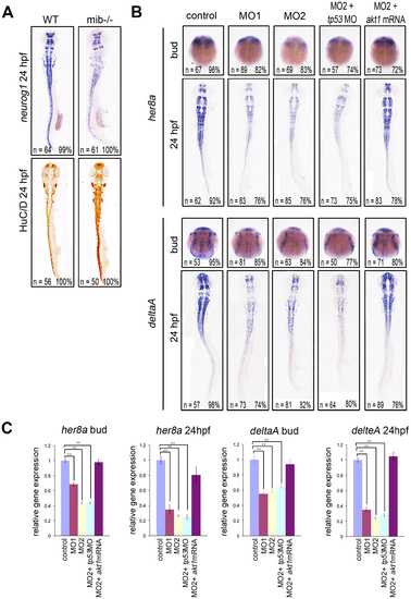
Akt1 is required for the expression of her8a and deltaA. (A) neurogenin1 expression was downregulated whereas HuC/D expression was upregulated in mibta52b mutants (right) compared with wild-type siblings (left). (B) In situ hybridization showed that her8a and deltaA were expressed at lower levels in akt1 morphants compared with the controls during the bud stage and at 24 hpf. This phenotype was restored by coinjection with akt1 mRNA but not with a tp53 morpholino. (C) The expression level in B was confirmed by qPCR. **, p<0.01. |
|
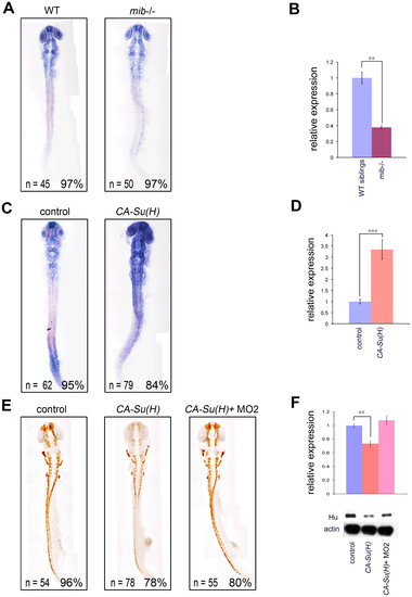
Notch-Su(H) signaling is sufficient to induce akt1 expression and inhibit neuronal differentiation. (A) akt1 expression was downregulated in Notch activity-deficient mind bomb mutant embryos compared with wild-type siblings as shown by in situ hybridization, which was confirmed by qPCR analysis in B. (C) akt1 expression levels were upregulated by constitutive-active Su(H) injection and quantified by qPCR in D. (E) Injection of constitutive-active Su(H) significantly downregulated HuC/D expression and this phenotype was nullified by akt1 knockdown. (F) Western blot analysis confirmed the levels of HuC/D expression shown in E. CA-Su(H), constitutive-active Su(H); **, p<0.01; ***, p<0.001. |
|
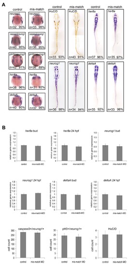
Embryos injected with a five-base mismatch morpholino show unaltered expression of neural markers. The specificity of the akt1 morpholinos was confirmed by injection with a morpholino with a five-base mismatch relative to MO2. Injection of this morpholino resulted in unaltered expression of neural markers showing by in situ hybridization (A) and quantitatively confirmed by qPCR (B). |

