- Title
-
Activated ALK Collaborates with MYCN in Neuroblastoma Pathogenesis
- Authors
- Zhu, S., Lee, J.S., Guo, F., Shin, J., Perez-Atayde, A.R., Kutok, J.L., Rodig, S.J., Neuberg, D.S., Helman, D., Feng, H., Stewart, R.A., Wang, W., George, R.E., Kanki, J.P., and Look, A.T.
- Source
- Full text @ Cancer Cell
|
Transgenic Gene Expression in the Sympathetic Neurons and the Interrenal Gland (A) Left: Schematic of a transverse section illustrating zebrafish anatomical structures, dorsal upward. Right: Schematic of a sagittal section illustrating zebrafish anatomical structures, anterior to left. (B) EGFP expression (green) in the zebrafish chain of sympathetic ganglia (arrowheads), the IRG (arrow), and medulla oblongata (asterisk) at 3 wpf. Lateral view of confocal-brightfield image, anterior to left. The magnified view of the boxed region is shown on the right. Scale bar represents 500 μm. (C) EGFP is coexpressed with TH in the SCG at 6 wpf (sagittal section); TH coexpression is indicated in red. Scale bar represents 20 μm. (D) EGFP is coexpressed with TH in the chain of sympathetic ganglia at 6 wpf (transverse section). TH coexpression is indicated in red. Scale bar represents 20 μm. (E) EGFP is coexpressed with TH in the IRG at 8 months postfertilization (mpf) (sagittal section). TH coexpression is indicated in red. Scale bar represents 100 μm. Ao, aorta; Cb, cerebellum; Chfn, chromaffin cells; E, esophagus; G, gill; H, heart; HK, head kidney; I, intestine; IRG, interregnal gland; KB, kidney body; L, liver; Me, medulla; Nc, notochord; SB, swim bladder; SC, spinal cord; SCG, superior cervical ganglion; Sym.C, sympathetic chain. See also Figure S1. EXPRESSION / LABELING:
|
|
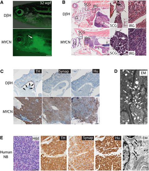
Neuroblastomas Arise in MYCN-Expressing Transgenic Zebrafish (A) Top: DβH fish. Bottom: MYCN fish with EGFP-expressing tumor (arrow) at 32 weeks postfertilization (wpf). Scale bar represents 1 mm. (B) Top: H&E-stained sagittal sections of DβH fish. Boxes indicate the SCG and the IRG, and are magnified in the right panels. Bottom: H&E-stained sagittal sections of MYCN fish with neuroblastic tumors. Boxes indicate the SCG and the IRG and are magnified in the right panels. Arrows indicate SCG neurons. The majority of tumors arise in the IRG of MYCN fish, although as seen in this example, tumor cells in the SCG were occasionally observed in individual fish that also had tumors in the IRG. G, gill; L, liver; I, intestine; IRG, interregnal gland; SCG, superior cervical ganglion; T, testis. Scale bars represent 50 μm. (C) Top: Sagittal sections through the interregnal gland of DβH fish. Chromaffin cells of the interregnal gland express TH (arrows). Bottom: Sagittal sections through the interregnal gland of a MYCN fish with EGFP-expressing tumor. Cells throughout the tumor in the interregnal gland express TH, Synaptophysin (Synap), and Hu. Scale bar represents 100 μm. (D) Electron microscopy (EM) reveals neurosecretory granules in the MYCN-expressing tumors (arrows). Scale bar represents 500 μm. (E) Pathological, immunohistochemical, and ultrastructural analyses of a human neuroblastoma. Arrows point to neurosecretory granules. Scale bars represent 500 μm (left panel), 100 μm (middle panels), and 2 µm (right panel), respectively. See also Figure S2. |
|
MYCN Expression Causes Sympathoadrenal Cell Loss (A) DβH transgenic line. Oblique views of dβh RNA expression (left panels); lateral views of EGFP expression in merged confocal-brightfield images (middle panels); dorsal views of th RNA expression (right panels). Arrows point to the SCG, and arrowheads point to the CG. Scale bar represents 100 μm. (B) MYCN transgenic line. MYCN expression causes loss of cells in the SCG (arrows). Scale bar represents 100 μm. (C) ALK transgenic line. ALK expression does not interfere with the SCG development (arrows). Scale bar represents 100 μm. (D) MYCN;ALK transgenic line. Loss of cells in the SCG is not rescued by activated ALK expression (arrows). Scale bar represents 100 μm. AAC, arch-associated catecholaminergic neurons; CG (arrowheads), cranial ganglia; DA, diencephalic dopaminergic neurons; e, ear; LC, locus coeruleus; MO, medulla oblongata; r, retina; SCG, superior cervical ganglion. See also Figure S4 and Table S1. EXPRESSION / LABELING:
PHENOTYPE:
|
|
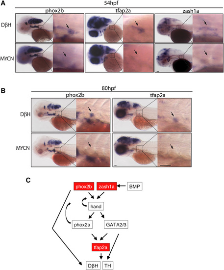
Expression of Early Sympathoadrenal Markers Is Absent in MYCN Transgenic Embryos during Early Development (A and B) Top panels: DβH; lower panels: MYCN transgenic fish. Expression of sympathoadrenal cell markers at 54 hpf (A) and 80 hpf (B). The magnified view of the boxed region is shown on the right. Arrows point to the superior cervical ganglion. Scale bars represent 50 μm (left panels) and 100 μm (right panels, magnified view). (C) Diagram of the genetic interactions of sympathoadrenal genes during early development. Arrows indicate the activation of target genes. Curved arrows indicate positive feedback regulation. See also Figure S5. EXPRESSION / LABELING:
|
|
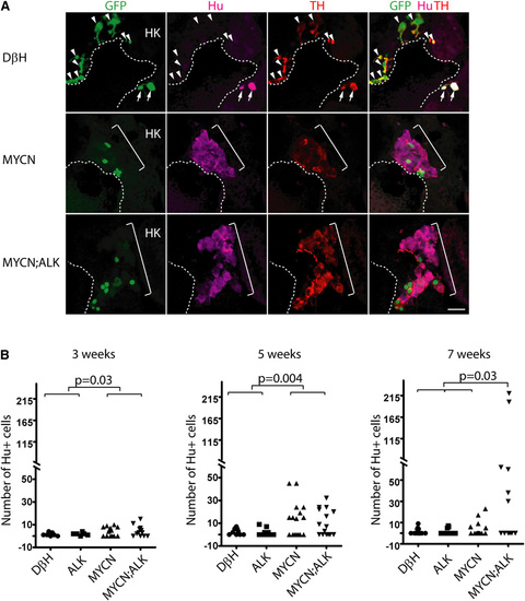
MYCN Causes Hu+ Cell Hyperplasia in the Interrenal Gland (A) Sagittal sections through the interrenal gland in DβH (top panels), MYCN (middle panels), and MYCN;ALK (lower panels) transgenic fish at 5wpf (dorsal up, anterior left). EGFP, green; Hu, magenta; TH, red. Representative sections through the interrenal gland in DβH fish contain three to five GFP+/Hu+/TH+ sympathetic neuroblasts (arrows) and many GFP+/Hu-/TH+ chromaffin cells (arrowheads). Hu+ cell numbers increase in MYCN and MYCN;ALK fish (brackets), and can be GFP+ and TH+. Dotted lines indicate the head kidney (HK) boundary. Scale bar represents 20 μm. (B) Numbers of Hu+ interrenal gland cells in DβH, ALK, MYCN, and MYCN;ALK transgenic fish at 3, 5, and 7 weeks. Means of Hu+ cell numbers were compared by the two-tailed Wilcoxon signed-rank test. See also Figure S6. EXPRESSION / LABELING:
PHENOTYPE:
|
|
ALK Inhibits a Developmentally-Timed Apoptotic Response Triggered by MYCN Overexpression in the Interrenal Gland (A) Numbers of Hu+ interrenal gland cells in the DβH, ALK, MYCN, and MYCN;ALK fish at 5.5 wpf. Means of Hu+ cell numbers were compared by the two-tailed Wilcoxon signed-rank test. (B) Numbers of apoptotic Hu+ interrenal gland cells in the DβH, ALK, MYCN, and MYCN;ALK fish at 5.5 wpf. The numbers of transgenic fish at 5.5 wpf with apoptotic Hu+ cells in the interrenal gland were compared by two-tailed Fisher exact test. (C) Sagittal sections through the interrenal gland in MYCN (top panels) and MYCN;ALK (bottom panels) transgenic fish at 5.5 wpf (dorsal up, anterior left). Hu, green; activated Caspase-3, red. Hu+, activated Caspase-3+ apoptotic cells were detected in the MYCN transgenic fish (arrowheads). Dotted lines indicate the head kidney (HK) boundary. Scale bars represent 10 μm. See also Figure S7. PHENOTYPE:
|

ZFIN is incorporating published figure images and captions as part of an ongoing project. Figures from some publications have not yet been curated, or are not available for display because of copyright restrictions. EXPRESSION / LABELING:
|
Reprinted from Cancer Cell, 21(3), Zhu, S., Lee, J.S., Guo, F., Shin, J., Perez-Atayde, A.R., Kutok, J.L., Rodig, S.J., Neuberg, D.S., Helman, D., Feng, H., Stewart, R.A., Wang, W., George, R.E., Kanki, J.P., and Look, A.T., Activated ALK Collaborates with MYCN in Neuroblastoma Pathogenesis, 362-373, Copyright (2012) with permission from Elsevier. Full text @ Cancer Cell


