- Title
-
HSPG-Deficient Zebrafish Uncovers Dental Aspect of Multiple Osteochondromas
- Authors
- Wiweger, M.I., Zhao, Z., van Merkesteyn, R.J., Roehl, H.H., and Hogendoornm, P.C.
- Source
- Full text @ PLoS One
|
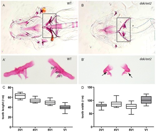
ext2-/- mutant displays severe tooth phenotype. Ventral view of alizarin-red-stained craniofacial skeleton and teeth at 6 dpf (A, B) and dissected and flat mounted 5th pharyngeal arches with teeth (A2, B2) reveals the presence on each pharyngeal arch of 3 teeth in siblings (A, A2) and only one misshapen tooth in ext2-/- larvae (B, B2). Note that the rod shaped branchial arch 5 to which the teeth attach is also ossified. Arrows point incomplete ossification of the mutant tooth. Tooth phenotype consisting of one misshapen tooth was observed in all (n>500) analysed ext2-/- embryos whereas heterozygote fish were indistinguishable from WT. Tooth lengths varies between 3V1, 4V1 and 5V1 in siblings (P<0.003). Each of those teeth was significantly longer then dak-tooth (P<0.0001) (C). Tooth widths of 3V1 and 5V1 were similar between siblings, and both were significantly narrower than 4V1 (D). ext2-/--tooth was significantly broader than any of the siblings teeth (3V1, P<0.0001; 4V1, P = 0.023 and 5V1, P = 0.0001) (D). White boxes, siblings; grey boxes, homozygote mutant. Scale bar = 0.1 mm. PHENOTYPE:
|
|
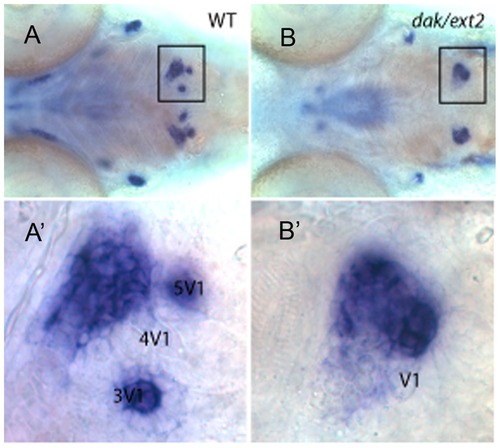
The attachment of the 1st tooth occurs on time in ext2-/- mutant. The expression of the osterix gene at 96 hpf underlines the pharyngeal arches in sibling (A, A2) and ext2-/- mutant (B, B2). At this stage, osterix is also expressed in the sibling in the tooth germs of 3V1 and 5V1, but lost in the 4V1. Mineralised dentine outlines the first to develop and attach – the 4V1 tooth. Note that single tooth that does not express osterix is also attached into osterix-positive pharyngeal arch in the ext2-/- mutant. A2 and B2 are higher magnification images of A and B. Scale bar = 0.1 mm. |
|
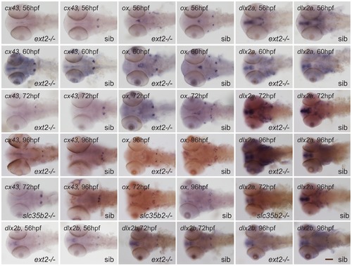
The expression pattern of the dental markers at three stages of development. A panel of markers was used to dissect the identity of the ext2-/--tooth (dak). Progress of tooth development was monitored at 56, 72 and 96 hpf using dlx2a, dlx2b and cx43 markers. slc35b2-/- (pic) mutant was included as it has 2 teeth located to the end of pharyngeal arch. Ossification of teeth in slc35b2-/- is also delayed and progresses only in one tooth. ext2-/- and slc35b2-/- mutants were sorted by phenotype. A, ventral view and B, dorsal view. Scale bar = 0.1 mm. |
|
Tooth phenotype in zebrafish mutants affected in FGF, IHH and PKC pathways. Pharyngeal arch together with teeth from 6-days-old WT and dackel (dak/ext2), detour (dtr/gli1), heart and soul (has/prkci), pinscher (pic/slc35b2) and you too (yot/gli2a) homozygote mutants were stained with alizarin red, dissected and flat mounted. The average length of the 5th arch in WT larvae is 150 μm. |
|
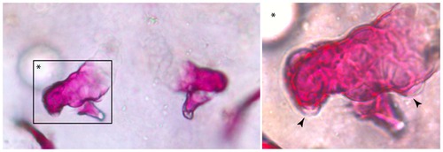
FGF8 stimulates growth of additional tooth-bud-like structures in ext2-/- mutant. Beads were implanted at 36–39 hpf on one side of the body into an area in between the heart, ear and pectoral fin, where the teeth start to form. At 5 dpf, fish were fixed and stained with Alizarin red. Tooth-buds-like structures were formed on the pharyngeal arch neighboured by FGF-coated bead (arrowhead). Opposite arch was not affected. The tooth-bud-like structures were observed on each side of ext2-/--tooth. Asterisk indicate position of the bead. |
|
Tooth organization in adult ext2(+/-) mutant fish. Adult dak heterozygote mutant has significantly less teeth in the dorsal row (A); whereas the number of teeth in the mediodorsal row (B) and ventral row (C) are similar to WT. Examples of pharyngeal teeth from adult ext2+/-: WT-like pharyngeal arch (D, D2), pharyngeal arch with a super-numeral tooth found in the ventral row (E, E2), and large gap between teeth (F, F2). D2, E2 and F2, larger views of D, E and F. Scale bars correspond to 0.1 mm. PHENOTYPE:
|
|
Dental defects are present in 20% of adult ext2+/- mutant fish. Lateral view of two ventral teeth stained with Alizarin red. In most cases, WT-like teeth were present (A, D). However, on few occasions we also observed: enamel malformation (B, E, F) or misshapen crowns (C, C2). Teeth start to calcify from the tip toward the base; hence the lack of staining at the base of 2V is most likely reflects uncompleted ossification of a recent replaced tooth – see black arrowhead (C). Teeth from MO patients (G, H). Note extra buckle in H (arrow head) which resembles split crown observed in ext2-/- fish. C2 is a higher magnification of C. White arrows indicate lesions. Scale bars correspond to 0.1 mm. PHENOTYPE:
|
|
Tooth ossification is delayed in ext2-/- mutant. Is indicated by Alizarin red stain at 96 hpf, single tooth is ossified in both ext2-/- mutant and its siblings. Interestingly, the ossification of the pharyngeal arches starts in the mid part in siblings (A, A2) and at the end of arch in ext2-/- mutant (B, B2). Moreover, weaker intensity of the Alizarin red in ext2-/- suggests general delay in ossification. A2 and B2, line outline of the branchial arch 5 and attached teeth. Scale bar = 0.1 mm. PHENOTYPE:
|
|
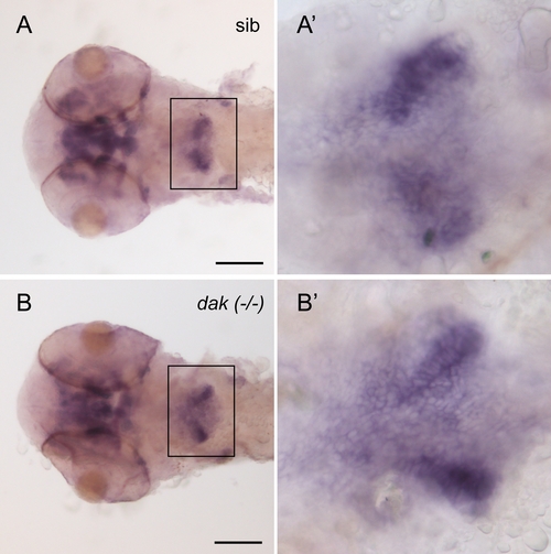
The expression pattern of pitx2 indicates slight defect in thickening of the pharyngeal epithelium in the ext2-/- mutant. pitx2-expressing bilateral domain in the ext2-/- are of similar length but more narrower than the one from siblings. A, siblings and B, ext2-/- at 56 hpf. A2 and B2, magnification of the pharyngeal area. Scale bar = 0.1 mm. |
|
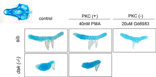
Inhibition of PKC affects tooth formation. Similarly to has (PKC) mutant, one tooth-phenotype was also observed in fish treated with PKC inhibitor. PMA – activator of PKC does not stimulate formation of additional teeth in WT, nor rescues tooth phenotype in the dak homozygote mutant. Cartilaginous skeletons were stained with Alcian blue at 6 dpf. Pharyngeal arches were dissected out and flat mounted. |
|
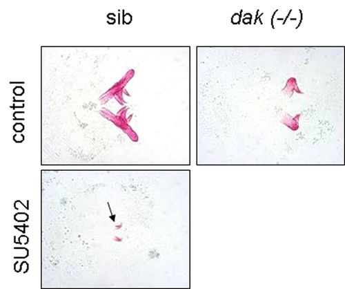
Inhibition of FGF by SU5402 tooth formation in WT and dak mutant. Embryos were treated from 50 hpf till 6 dpf. The ext2-/- fish treated with SU5402 does not form teeth hence picture was not included. Alizarin-red-stained pharyngeal arches were dissected and flat mounted. Arrow points a single bilateral tooth formed in dak siblings. |
|
Tooth morphology in adult dak heterozygote mutant. Cross section of teeth from adult fish did not reveal any obvious morphological differences between WT and ext2-/-. 4 μm sections of teeth were stained with haematoxylin and eosin. |
|
Accumulation of cells undergoing cell death at the end of the pharyngeal arch in the ext2-/- mutant. TUNEL staining was performed in fish at 6 dpf. Pharyngeal arches were dissected and flat mounted. PHENOTYPE:
|

