- Title
-
Wdr18 Is Required for Kupffer's Vesicle Formation and Regulation of Body Asymmetry in Zebrafish
- Authors
- Gao, W., Xu, L., Guan, R., Liu, X., Han, Y., Wu, Q., Xiao, Y., Qi, F., Zhu, Z., Lin, S., and Zhang, B.
- Source
- Full text @ PLoS One
|
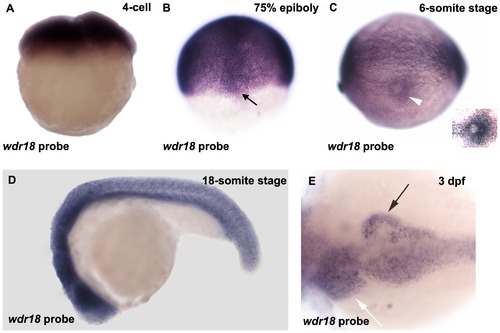
wdr18 expression pattern during early embryonic development in zebrafish. (A) wdr18 is maternally expressed. (B) Expression pattern of wdr18 at the 75%-epiboly stage. Black arrow indicates the staining of dorsal forerunner cells (DFCs). (C) wdr18 expression is detected around the Kupffer′s vesicle (white arrowhead) at the 6-somite stage. Inset is an overexposed magnified image showing elevated wdr18 expression around the KV. (D) wdr18 is detected ubiquitously throughout the whole embryo at 18-somite stage. (E) At 3 dpf, wdr18 is expressed in endodermal organs, including the exocrine pancreas (black arrow), liver (white arrow), and intestine. EXPRESSION / LABELING:
|
|
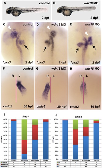
Knockdown of wdr18 disturbed L-R asymmetry development of internal organs. (A, B) The overall appearance of a wdr18 morphant embryo (B) is comparable to a wildtype control embryo (A). (C) Expression of foxa3, a marker for the endoderm, showed that the liver (white arrowheads) and pancreas (black arrows) were positioned to the left and the right of the midline in a control embryo at 2 dpf, respectively. In morphant embryos, the positions of the liver and pancreas were reversed (D), or the pancreas appeared duplicated (two black arrows) when the liver was randomized (E). cmlc2, a marker for the heart, was examined in embryos at 30 hpf. After the heart tube formation, leftward bending of the heart was observed in control embryos (F). By contrast, wdr18 morphant embryos exhibited randomized movements: leftward, rightward (G), or no movement (H). The proportion of each phenotype is summarized in (I) and (J). Dup-pa in (I) indicates embryos with duplicated pancreas. M, R, and L in (J) stand for middle, right, and left, respectively. Injection of 100 pg wdr18 mRNA partially rescued the endodermal organ phenotype. The statistical results are summarized in (I) and (J). (C–E) Dorsal views, anterior to the top. (F–H) Ventral views, anterior to the top. PHENOTYPE:
|
|
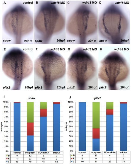
wdr18 morpholino caused randomized expression of spaw and pitx2. In nearly all control embryos, spaw and pitx2 showed left-sided expression (A, E), whereas wdr18 morphants showed left-sided (B, F), right-sided (C, G), or bilateral (D, H) spaw or pitx2 expression in the LPM. (I, J) The percentage of control, morphant, mRNA-rescued and mRNA over-expressed embryos with left-sided (L), right-sided (R), or bilateral (Bi) spaw and pitx2 expression. (A–H) Dorsal views, anterior to the top. EXPRESSION / LABELING:
PHENOTYPE:
|
|
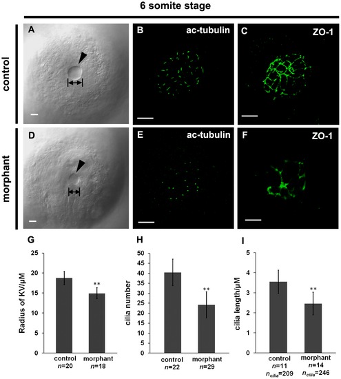
KV formation is disturbed in wdr18 morphant embryos. (A, D) DIC images of KV in control (A) and wdr18 morphant (D) embryos. Note the KV size in control was larger than that in the morphants. (B, E) Confocal images of cilia inside KV in control (B) and morphant (E) embryos detected by a fluorescent anti-acetylated tubulin antibody. Shown is a 3D projection of multiple focal slices spanning KV at an interval of 0.4 µm. (C, F) anti-ZO-1 staining of the KV in 6- to 8-somite stage embryos. The control embryo (C) developed a KV with a large fluid-filled lumen, showing a uniform ZO-1-positive tight junction lattice within the lining of the KV, while the wdr18 morphant embryo (F) showed a dysmorphic lattice of ZO-1 labeling within the KV. (G) Measurement of the radius of individual KV in control and morphant embryos at 6- to 8-somite stage and statistical analyses. (H) Graphic representation of quantification results of cilia number per KV in both control and morphant embryos at 6- to 8-somite stage. (I) Quantification result of cilia length in control and wdr18 morphant embryos at 6- to 8-somite stage. The double asterisks in G, H and I indicate that the difference is extremely significant between experimental and control samples (P<0.0001, Student′s t-test). Scale bars: 20 µm in A and D; 15 µm in B, C, E and F. PHENOTYPE:
|
|
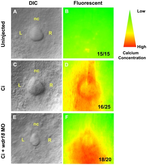
Correct Ca2+ distribution in the KV during early somite stages requires wdr18. (A–F) Embryos were injected at the 1-cell stage with Ca2+ indicator (Ci) or co-injected with wdr18 MO1. Ca2+ patterns at the 5- to 8-somite stage were imaged by fluorescence microscopy and the images were converted to an intensity scale (colors from red to green indicate intensity from high to low). (A, B) The autofluorescent image of an un-injected control embryo (B) with its corresponding DIC image (A). (C, D) A control embryo injected with Ca2+ indicator only, showing fluorescence in the KV with elevated signals on the left side. (E, F) An embryo co-injected with Ca2+ indicator and wdr18 MO1. The KV is smaller and the fluorescence signal is evenly distributed throughout the KV. nc: notochord. The ratio shown in the lower right corner of B, D and F indicate the number of defective embryos versus total embryos. PHENOTYPE:
|
|
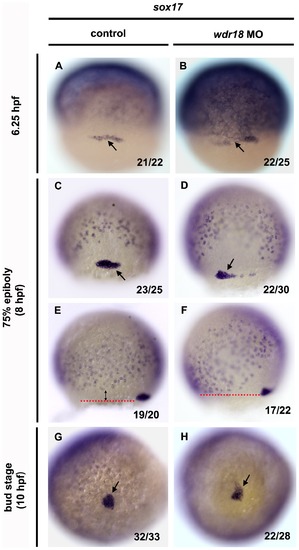
DFC migration is affected in wdr18 morphant embryos. (A–H) In situ hybridization with sox17 showing the migration and clustering process of DFCs in control and wdr18 morphant embryos at 6.25 hpf, 75%-epiboly stage and bud stage. sox17 is a marker of endodermal cells and DFCs. The pepper-like staining represents endodermal cells and the grouped cells (black arrow) indicate DFCs. (A, C, G) The DFCs in control embryos first appeared as a horizontal line of cells (A), and then migrated toward the vegetal pole while forming a compact oval-shaped cluster (C) and eventually a round condensed cell mass at the end of epiboly, which will gradually differentiate into the Kupffer′s vesicle. (B, D, H) In wdr18 morphant embryos, the shape of DFCs looked nearly the same as control embryos at 6.25 hpf (B), however, the DFCs failed to form a single compact cell cluster at 75%-epiboly stage (D). Although the DFCs in morphant embryos could finish clustering process at the end of epiboly, the cell mass was frequently appeared misshaped (H). (E, F) The location of the DFCs relative to the endodermal cell layer. (E) In a control embryo, the DFC migratory front (red dotted line) is further ahead of the endodermal cell layer as a result of DFC migration towards the vegetal pole. (F) In the morphant embryo, the DFC migratory front is almost at the same level as the endodermal layer, indicating that DFC migration is slower and uncoupled with overall gastrulation movements. The ratio shown in the lower right corner of each image indicate the number of defective embryos versus total embryos. (A–D) Dorsal views, animal pole to the top. (E, F) Side views, dorsal to the right, animal pole to the top. PHENOTYPE:
|
|
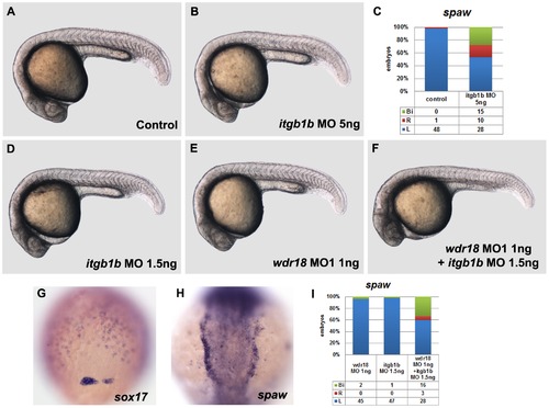
wdr18 and integrin β1b act genetically in the regulation of laterality development. (A) A control embryo under a light microscope. (B) The overall morphology of embryos injected with 5 ng of itgb1b morpholino is indistinguishable from controls. (C) The percentage of control and itgb1b morphant embryos with left-sided (L), right-sided (R), or bilateral (Bi) spaw expression. (D, E, F) The morphology of embryos injected with 1.5 ng itgb1b morpholino (D) or 1 ng wdr18 MO1 (E), and embryos injected with a combination of 1 ng wdr18 MO1 and 1.5 ng itgb1b morpholino (F) look relatively normal, except for a minor difference in body curvature (F). (G) sox17 staining in embryos injected with a combination of 1 ng wdr18 MO1 and 1.5 ng itgb1b morpholino indicates that DFC migration is affected. Dorsal view, animal pole to the top. (H) An example of bilateral spaw expression in the LPM of embryos injected with a combination of 1 ng wdr18 MO1 and 1.5 ng itgb1b morpholino. Dorsal view, anterior to the top. (I) The percentage of control embryos injected with 1.5 ng itgb1b morpholino, 1ng wdr18 MO1, or a combination of 1 ng wdr18 MO1 and 1.5 ng itgb1b morpholino showing left-sided (L), right-sided (R), or bilateral (Bi) spaw expression. PHENOTYPE:
|
|
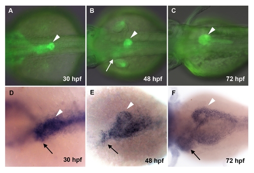
GFP fluorescent signal in mp235b embryos and wdr18 expression in endodermal organs. (A–C) GFP expression pattern in mp235b transgenic line. (D–F) Whole-mount in situ hybridization showing wdr18 gene expression in endodermal region during the first 3 days of development. Arrowheads indicate pancreas and arrows indicate liver region. All the embryos were shown as dorsal views with anterior to the left. |
|
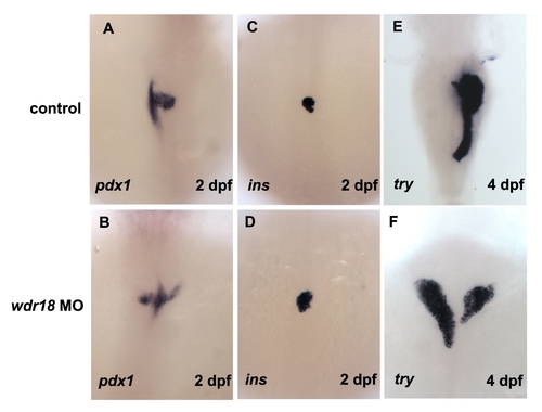
The effect of wdr18 knockdown on endodermal organs. (A, B) pdx1 was used as a marker for the pancreatic bud. In morphant embryos, the pancreas appeared on both sides of the body (B), while in control embryos it resided on the right side. (C, D) insulin (ins) was used as a differentiation marker for pancreatic β cells. Note that the development of β cells was not affected after wdr18 morpholino injection. (E, F) In situ hybridization of trypsin (try) revealing the exocrine pancreas at 4 dpf. In morphant embryos, the exocrine pancreas differentiated normally, but appeared in duplicated form. Dorsal views, anterior to the top. |
|
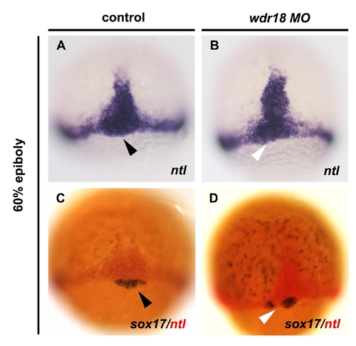
Knockdown of wdr18 led to defects in DFC migration without affecting DFC specification. (A) In situ hybridization result of ntl, showing the notochord precursor cells at the midline, marginal cells, and DFCs (black arrowhead) in a control embryo at 60%-epiboly stage. (B) The midline structure and marginal cells were not affected, while the DFC region, marked by a white arrowhead, seemed reduced or mis-localized in a wdr18 morphant. (C, D) Double in situ hybridization result showed clear staining of both ntl (red) and sox17 (blue) positive cells in wdr18 morphant embryos, indicating DFCs were still present after knockdown of wdr18, although they were not properly organized as a single cluster. Dorsal views, animal pole to the top. |
|
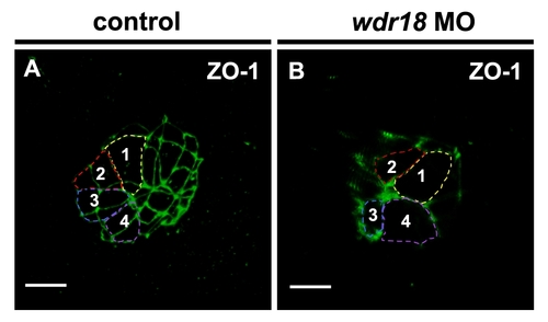
Knockdown of wdr18 did not change the size of cells composing Kupffer′s vesicle. 3D reconstruction confocal images of the KV in control and wdr18 morphant embryos revealed by immuno-staining with ZO-1 antibody at 6-somite stage. For the convenience of outlining the cell shape, the 3D images were rotated to the proper position until the boundaries of the marked cells was clearly seen. (A) In a control embryo, the cell connection was obvious and we outlined four cells with different colors and numbered them from 1 to 4. (B) In a wdr18 morphant, we similarly outlined and numbered four cells for comparison. The size of the marked cells showed no obvious difference comparing with that in control embryos. Scale bars: 15 μm. |

