
Fig. S5
- ID
- ZDB-IMAGE-110920-12
- Publication
- Gao et al., 2011 - Wdr18 Is Required for Kupffer's Vesicle Formation and Regulation of Body Asymmetry in Zebrafish
- All Figures
- Figures for Gao et al., 2011
|
Fig. S5
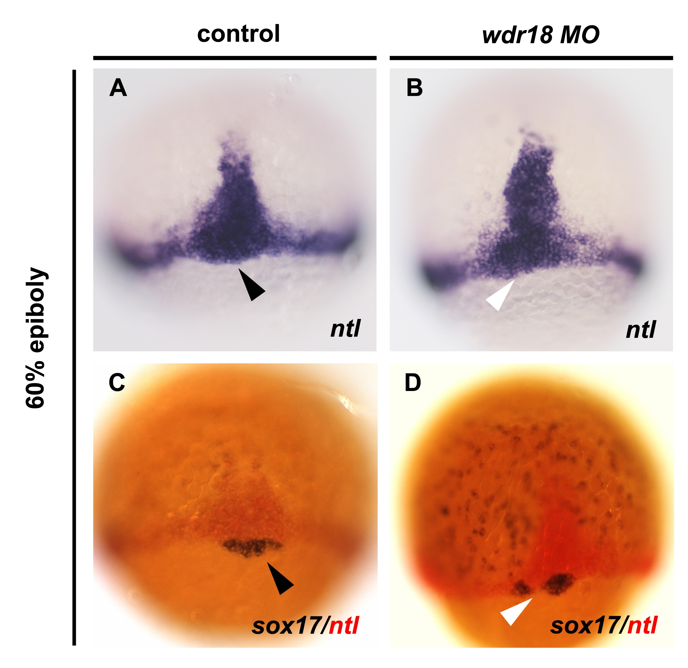
Knockdown of wdr18 led to defects in DFC migration without affecting DFC specification. (A) In situ hybridization result of ntl, showing the notochord precursor cells at the midline, marginal cells, and DFCs (black arrowhead) in a control embryo at 60%-epiboly stage. (B) The midline structure and marginal cells were not affected, while the DFC region, marked by a white arrowhead, seemed reduced or mis-localized in a wdr18 morphant. (C, D) Double in situ hybridization result showed clear staining of both ntl (red) and sox17 (blue) positive cells in wdr18 morphant embryos, indicating DFCs were still present after knockdown of wdr18, although they were not properly organized as a single cluster. Dorsal views, animal pole to the top.