
Fig. 6
- ID
- ZDB-IMAGE-110920-9
- Genes
- Publication
- Gao et al., 2011 - Wdr18 Is Required for Kupffer's Vesicle Formation and Regulation of Body Asymmetry in Zebrafish
- All Figures
- Figures for Gao et al., 2011
|
Fig. 6
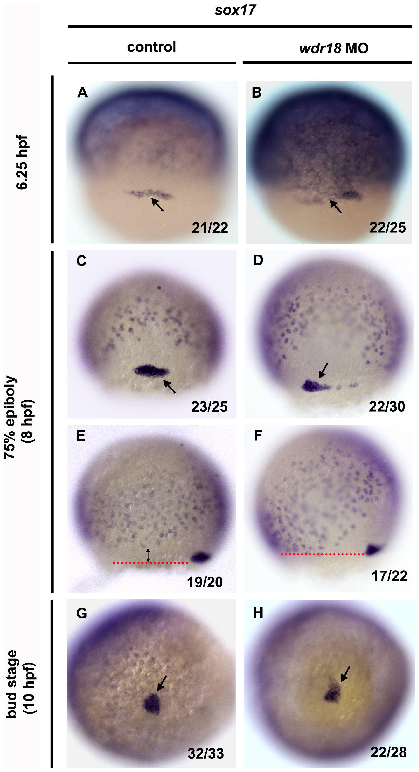
DFC migration is affected in wdr18 morphant embryos.
(A–H) In situ hybridization with sox17 showing the migration and clustering process of DFCs in control and wdr18 morphant embryos at 6.25 hpf, 75%-epiboly stage and bud stage. sox17 is a marker of endodermal cells and DFCs. The pepper-like staining represents endodermal cells and the grouped cells (black arrow) indicate DFCs. (A, C, G) The DFCs in control embryos first appeared as a horizontal line of cells (A), and then migrated toward the vegetal pole while forming a compact oval-shaped cluster (C) and eventually a round condensed cell mass at the end of epiboly, which will gradually differentiate into the Kupffer′s vesicle. (B, D, H) In wdr18 morphant embryos, the shape of DFCs looked nearly the same as control embryos at 6.25 hpf (B), however, the DFCs failed to form a single compact cell cluster at 75%-epiboly stage (D). Although the DFCs in morphant embryos could finish clustering process at the end of epiboly, the cell mass was frequently appeared misshaped (H). (E, F) The location of the DFCs relative to the endodermal cell layer. (E) In a control embryo, the DFC migratory front (red dotted line) is further ahead of the endodermal cell layer as a result of DFC migration towards the vegetal pole. (F) In the morphant embryo, the DFC migratory front is almost at the same level as the endodermal layer, indicating that DFC migration is slower and uncoupled with overall gastrulation movements. The ratio shown in the lower right corner of each image indicate the number of defective embryos versus total embryos. (A–D) Dorsal views, animal pole to the top. (E, F) Side views, dorsal to the right, animal pole to the top.