
- Title
-
Zebrafish monosex population reveals female dominance in sex determination and earliest events of gonad differentiation
- Authors
- Tong, S.K., Hsu, H.J., and Chung, B.C.
- Source
- Full text @ Dev. Biol.
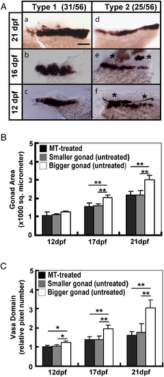
Comparison of the gonad size of MT-treated and wildtype juveniles. (A) Whole mount in situ RNA hybridization of vasa in two typical sets of gonads. Examples of the type 1 with fat gonads (a, b, c) and type 2 with slender gonads (d, e, f) at larvae of 21, 16 and 12 dpf are shown. The scale bar represents 100 μm. (B) Areas of gonads and (C) expression domains of vasa from MT-treated and two groups of wildtype juveniles are compared at 12, 17, and 21 dpf. The average intensity of 12-dpf MT-treated gonads is taken as 100. The number in parentheses in (A) indicates the number of gonads with a typical shape versus the total number of gonads. Asterisks in (A) demonstrate the pigment near the gonads. *P < 0.05, **P < 0.01. EXPRESSION / LABELING:
|
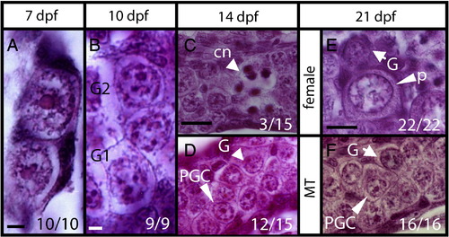
Histology of developing gonads. Longitudinal sections of gonads at (A) 7 dpf, (B) 10 dpf, (C,D) 14 dpf, and (E,F) 21 dpf. (A) Gonads contain primordial germ cells (PGC) with a single large round nucleolus. (B) Some germ cells differentiated into gonocytes 1 and 2 (G1, G2) with multiple nucleoli in the big rounded nucleus at 10 dpf. (C) Some gonads (3/15) with differentiated stage-I meiotic oocytes showing condensed chromatin (cn) at 14 dpf. (D) Other gonads (12/15) contain only PGCs and gonocytes. (E) Differentiating ovary with perinucleolar oocytes (p), and gonocytes (G). (F) MT-treated gonad with undifferentiated germ cells of gonocyte (G) or PGC morphology. Scale bars represent 5 μm in (A–B), 20 μm in (C–F). Numbers in the corner indicate the number of larvae with a typical RNA expression pattern versus the total number of larvae. |
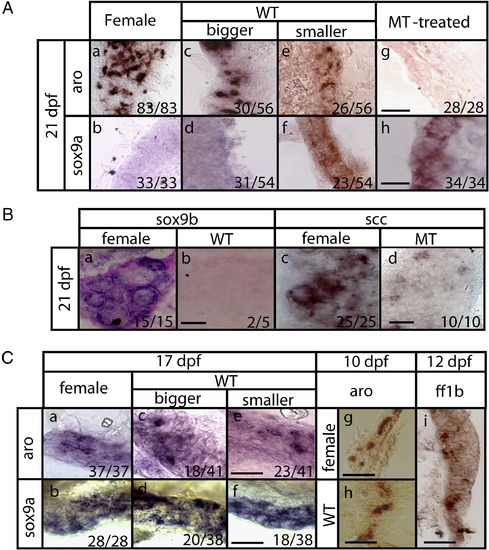
Expression of gonad markers in testes and ovaries at different stages. Expression of sox9a, aro and ff1b in (A) 21-dpf whole-mount (B) 21-dpf cross-sectioned and (C) 17-, 10-, and 12-dfp whole-mount gonads was detected by in situ RNA hybridization. Positive signals are shown in blue or brown color. All sectioned slides were counter-stained with Nuclear Fast Red. All female fish are progeny of super mothers. WT: wildtype fish. Bigger and smaller gonads in (A) and (C) refer to gonad sizes. Numbers at the corner indicate the number of larvae with a typical RNA expression pattern versus the total number of larvae. Scale bars represent 40 μm in (A and Ca–f), 20 μm in (B) and 25 μm in (Cg–i). aro: cyp19a1a, ff1b: nr5a1a, scc: cyp11a1. EXPRESSION / LABELING:
|

Lack of sox9a expression in a 10-dpf gonad. Whole-mount in situ hybridization of (A) vasa and (B) Sox9a in a 10 day-old gonad. EXPRESSION / LABELING:
|
Reprinted from Developmental Biology, 344(2), Tong, S.K., Hsu, H.J., and Chung, B.C., Zebrafish monosex population reveals female dominance in sex determination and earliest events of gonad differentiation, 849-856, Copyright (2010) with permission from Elsevier. Full text @ Dev. Biol.