
Fig. 3
- ID
- ZDB-FIG-100816-22
- Publication
- Tong et al., 2010 - Zebrafish monosex population reveals female dominance in sex determination and earliest events of gonad differentiation
- Other Figures
- All Figure Page
- Back to All Figure Page
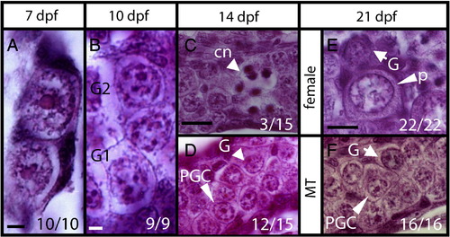
Histology of developing gonads. Longitudinal sections of gonads at (A) 7 dpf, (B) 10 dpf, (C,D) 14 dpf, and (E,F) 21 dpf. (A) Gonads contain primordial germ cells (PGC) with a single large round nucleolus. (B) Some germ cells differentiated into gonocytes 1 and 2 (G1, G2) with multiple nucleoli in the big rounded nucleus at 10 dpf. (C) Some gonads (3/15) with differentiated stage-I meiotic oocytes showing condensed chromatin (cn) at 14 dpf. (D) Other gonads (12/15) contain only PGCs and gonocytes. (E) Differentiating ovary with perinucleolar oocytes (p), and gonocytes (G). (F) MT-treated gonad with undifferentiated germ cells of gonocyte (G) or PGC morphology. Scale bars represent 5 μm in (A–B), 20 μm in (C–F). Numbers in the corner indicate the number of larvae with a typical RNA expression pattern versus the total number of larvae. |
Reprinted from Developmental Biology, 344(2), Tong, S.K., Hsu, H.J., and Chung, B.C., Zebrafish monosex population reveals female dominance in sex determination and earliest events of gonad differentiation, 849-856, Copyright (2010) with permission from Elsevier. Full text @ Dev. Biol.