
|
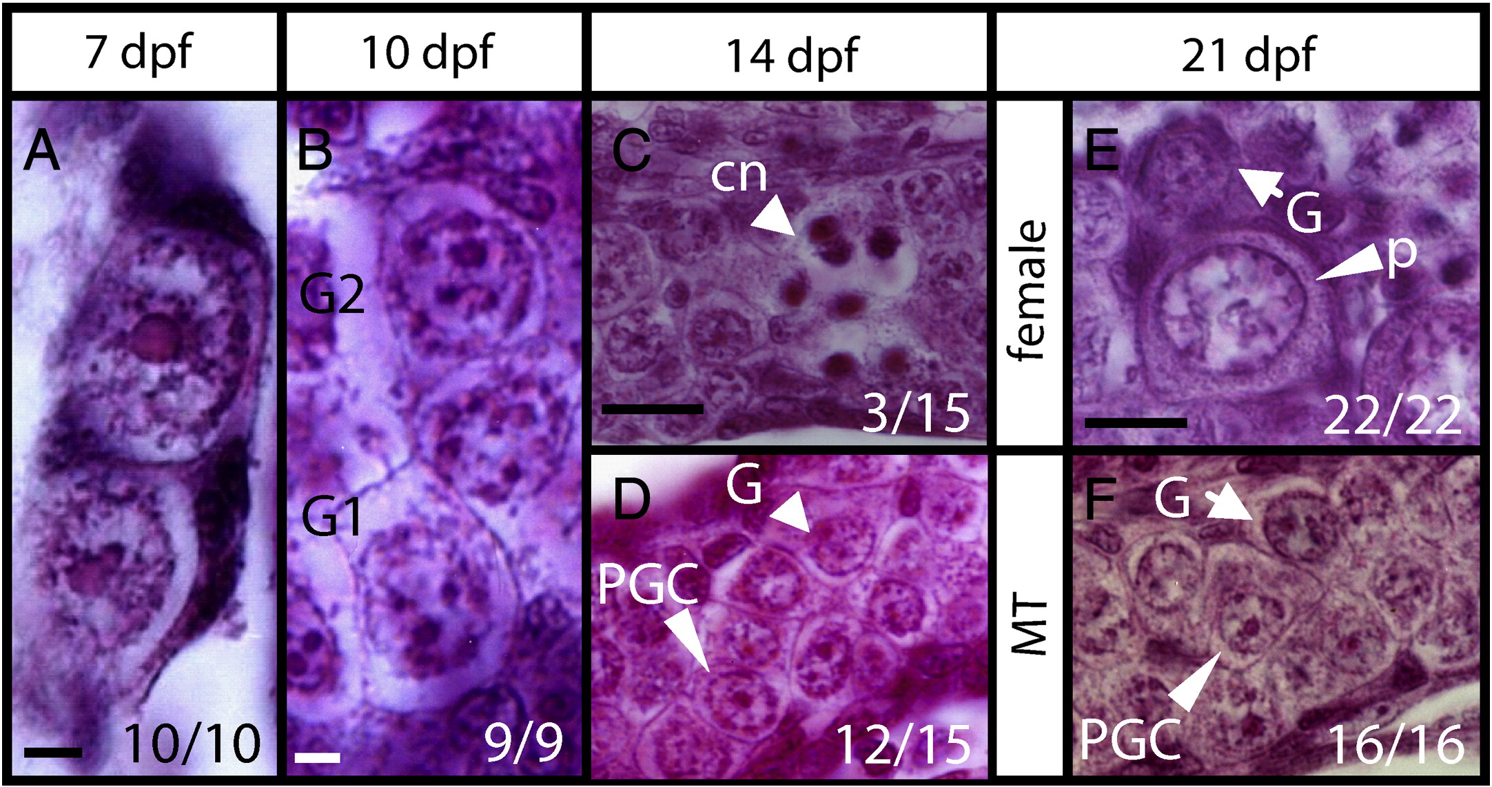
Fig. 3 Histology of developing gonads. Longitudinal sections of gonads at (A) 7 dpf, (B) 10 dpf, (C,D) 14 dpf, and (E,F) 21 dpf. (A) Gonads contain primordial germ cells (PGC) with a single large round nucleolus. (B) Some germ cells differentiated into gonocytes 1 and 2 (G1, G2) with multiple nucleoli in the big rounded nucleus at 10 dpf. (C) Some gonads (3/15) with differentiated stage-I meiotic oocytes showing condensed chromatin (cn) at 14 dpf. (D) Other gonads (12/15) contain only PGCs and gonocytes. (E) Differentiating ovary with perinucleolar oocytes (p), and gonocytes (G). (F) MT-treated gonad with undifferentiated germ cells of gonocyte (G) or PGC morphology. Scale bars represent 5 μm in (A–B), 20 μm in (C–F). Numbers in the corner indicate the number of larvae with a typical RNA expression pattern versus the total number of larvae.
Reprinted from Developmental Biology, 344(2), Tong, S.K., Hsu, H.J., and Chung, B.C., Zebrafish monosex population reveals female dominance in sex determination and earliest events of gonad differentiation, 849-856, Copyright (2010) with permission from Elsevier. Full text @ Dev. Biol.