
- Title
-
Suppression of the endoplasmic reticulum calcium pump during zebrafish gastrulation affects left-right asymmetry of the heart and brain
- Authors
- Kreiling, J.A., Balantac, Z.L., Crawford, A.R., Ren, Y., Toure, J., Zchut, S., Kochilas, L., and Creton, R.
- Source
- Full text @ Mech. Dev.

Cytosolic Ca2+ patterns in gastrula stage embryos. (A–C) Ratiometric imaging of cytosolic Ca2+ patterns at 6 hpf during normal development. (D–F) Cytosolic Ca2+ patterns at 6 hpf following inhibition of the ER Ca2+ pump with 0.5 μM thapsigargin from 4 to 6 hpf. Ca2+ concentrations are elevated in all embryonic regions, including the ventral margin (v), dorsal margin (d), and dorsal forerunner cells (DFC). (A and D) Overlay of bright field (grey-scale), the calcium indicator Oregon Green BAPTA-1 dextran (green), and the calcium-insensitive control Texas Red dextran (red). (B and E) Overlay of Oregon Green BAPTA-1 dextran and Texas Red dextran. (C and F) Pseudocolored ratiometric images (green/red). (G) The bar graph shows average cytosolic Ca2+ concentrations in the ventral margin (V), dorsal margin (D), and dorsal forerunner cells (DFC). The average Ca2+ concentration was measured in a circular region of interest with a diameter of 40 μm (ventral and dorsal margin) or 20 μm (dorsal forerunner cells). In the DMSO-treated controls, Ca2+ concentrations are significantly lower in the dorsal margin and DFC compared to the ventral margin (two-tailed t-test, P < 0.05, n = 13). Ca2+ concentrations increase significantly in the ventral margin, dorsal margin, and dorsal forerunner cells following treatment with 0.5 μM thapsigargin from 4 to 6 hpf (two-tailed t-test, P < 0.05, n = 13). Scale bar = 200 μm. |

Suppression of the ER Ca2+ pump during gastrulation affects heart laterality. (A) Control embryos, treated with 1 μl/ml DMSO from 4 to 6 hpf, and imaged at 30 and 50 hpf. The heart is positioned on the left at 30 hpf and shows a D-loop at 50 hpf, with the atrium (a) located on the left and the ventricle (v) on the right. (B–D) Embryos treated with 0.5 μM thapsigargin from 4 to 6 hpf and imaged at 30 and 50 hpf. Thapsigargin-treated embryos display a randomized cardiac orientation, including a reversed orientation or L-loop (B) a normal orientation or D-loop (C), and a centralized orientation or no-loop (D). Each embryo is shown in a bright field image (i, iv), pseudo-colored subtractive image (ii, v), and overlay of the bright field image and subtractive image (iii, vi). The images are shown as collected on an inverted microscope, with embryo’s left on the left side of the image, and the embryo’s right on the right side of the image. Scale bar = 200 μm. |

Suppression of the ER Ca2+ pump affects left–right laterality in the brain and gut. (A) Side view of pitx2 expression at the 22–25 somite stage (20 hpf). pitx2 is expressed in ventral and dorsal regions of the diencephalon. (B) Rostral view of a control embryo, treated with DMSO from 4 to 6 hpf, showing left-sided pitx2a expression in the dorsal diencephalon. (C and D) Embryos treated with 0.5 μM thapsigargin from 4 to 6 hpf frequently display right-sided (C) or bilateral (D) pitx2a expression in the dorsal diencephalon. The arrowheads point to pitx2a expression in the dorsal diencephalon. (E) Overlay of bright field image (grey-scale) and immunolabeling of neural patterns (green) in a DMSO-treated control embryo at 4 days post fertilization. (F) The prominent habenular nucleus was located on the left side in all DMSO-treated controls (n = 15). (G) Inhibition of the ER Ca2+ pump during gastrulation induces a left–right reversal of the prominent habenular nucleus in 39% of the embryos (n = 49). Neural patterns were labeled with an acetylated tubulin antibody and were imaged by confocal microscopy. (H) DMSO-treated control embryo at 3 dpf with a left-sided gut. (I) Thapsigargin treatment induces a reversal of gut laterality in 41% of the embryos (n = 41). The gut is stained in vivo by methylene blue in the culture medium. L, left habenular nucleus; R, right habenular nucleus. Panels B–D and E–G are oriented with embryo’s left on the left side of the image. Scale bar = 50 μm. EXPRESSION / LABELING:
|
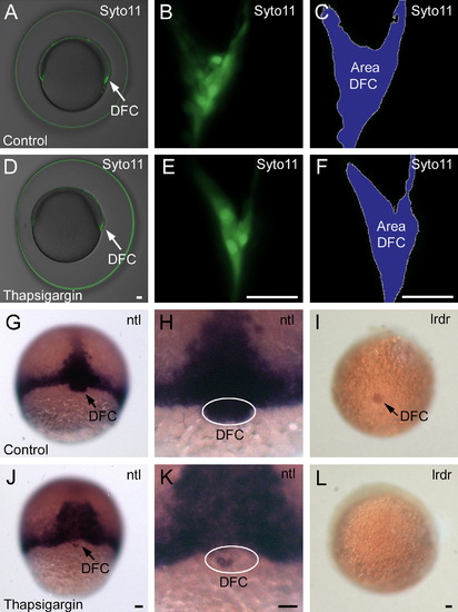
Ca2+-sensitive gene expression in the dorsal forerunner cells. (A–F) Syto11 labeling in the dorsal forerunner cells (DFC) at 7 hpf. The dorsal forerunner cells are present in the same location in the DMSO-treated control embryos (A–C), as they are in the thapsigargin-treated embryos (D–F). (G–L) ntl and lrdr RNA in situ hybridizations. DMSO-treated control embryos express ntl in the margin, notochord, and dorsal forerunner cells at 7 hpf (G and H) and express lrdr in the dorsal forerunner cells at 8 hpf (I). Thapsigargin treatment resulted in a strong down-regulation of ntl expression in the dorsal forerunner cells (J and K) and led to a complete inhibition of lrdr expression (L). Scale bar = 50 μm. EXPRESSION / LABELING:
|

Ca2+ manipulation affects the formation of Kupffer’s vesicle. (A and B) Control embryo treated with 1 μl/ml DMSO from 6 to 8 hpf, shown in lateral view (A) and posterior view (B) at the 9 somite stage (13 hpf). The arrows indicate the position of Kupffer’s vesicle. (C) The cilia in Kupffer’s vesicle were labeled by immunocytochemistry using an acetylated tubulin antibody. The vesicle shown in C is from a DMSO-treated control embryo and contains 70 cilia. (D and E) Embryo treated with 0.5 μM thapsigargin from 6 to 8 hpf and shown in a side view (D) and posterior view (E) at the 9 somite stage. Kupffer’s vesicle is absent. (F) Thapsigargin-treated embryo without a vesicle, which fails to develop cilia in the tail region. (G) Bar graph showing the number of embryos without a vesicle. (H) Embryos containing a vesicle were separated from embryos without a vesicle at 12–13 hpf and the two groups were examined for heart laterality defects at 50 hpf. Scale bars are 50 μm. |
Reprinted from Mechanisms of Development, 125(5-6), Kreiling, J.A., Balantac, Z.L., Crawford, A.R., Ren, Y., Toure, J., Zchut, S., Kochilas, L., and Creton, R., Suppression of the endoplasmic reticulum calcium pump during zebrafish gastrulation affects left-right asymmetry of the heart and brain, 396-410, Copyright (2008) with permission from Elsevier. Full text @ Mech. Dev.