
|
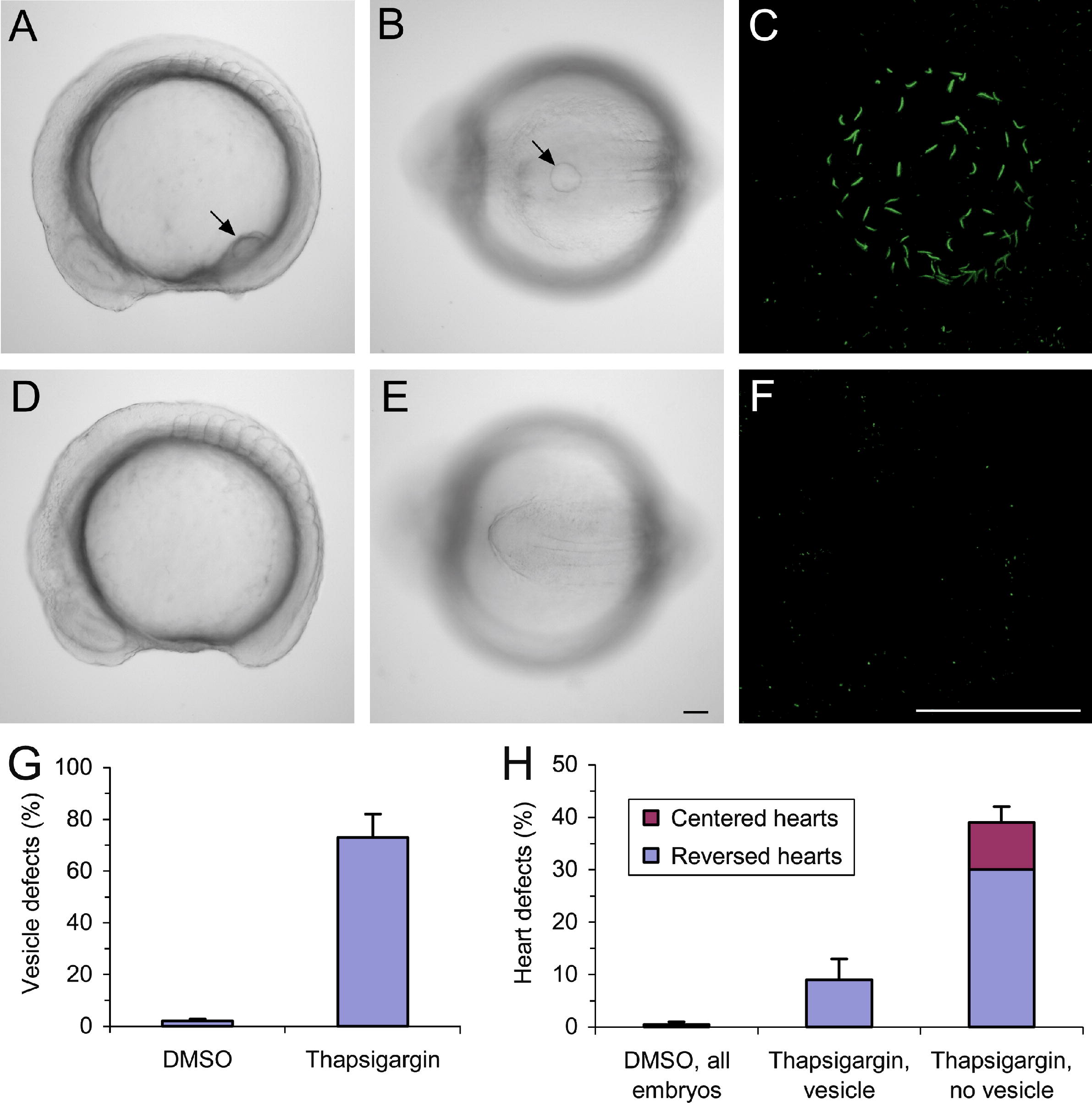
Fig. 6 Ca2+ manipulation affects the formation of Kupffer’s vesicle. (A and B) Control embryo treated with 1 μl/ml DMSO from 6 to 8 hpf, shown in lateral view (A) and posterior view (B) at the 9 somite stage (13 hpf). The arrows indicate the position of Kupffer’s vesicle. (C) The cilia in Kupffer’s vesicle were labeled by immunocytochemistry using an acetylated tubulin antibody. The vesicle shown in C is from a DMSO-treated control embryo and contains 70 cilia. (D and E) Embryo treated with 0.5 μM thapsigargin from 6 to 8 hpf and shown in a side view (D) and posterior view (E) at the 9 somite stage. Kupffer’s vesicle is absent. (F) Thapsigargin-treated embryo without a vesicle, which fails to develop cilia in the tail region. (G) Bar graph showing the number of embryos without a vesicle. (H) Embryos containing a vesicle were separated from embryos without a vesicle at 12–13 hpf and the two groups were examined for heart laterality defects at 50 hpf. Scale bars are 50 μm.
Reprinted from Mechanisms of Development, 125(5-6), Kreiling, J.A., Balantac, Z.L., Crawford, A.R., Ren, Y., Toure, J., Zchut, S., Kochilas, L., and Creton, R., Suppression of the endoplasmic reticulum calcium pump during zebrafish gastrulation affects left-right asymmetry of the heart and brain, 396-410, Copyright (2008) with permission from Elsevier. Full text @ Mech. Dev.