
|
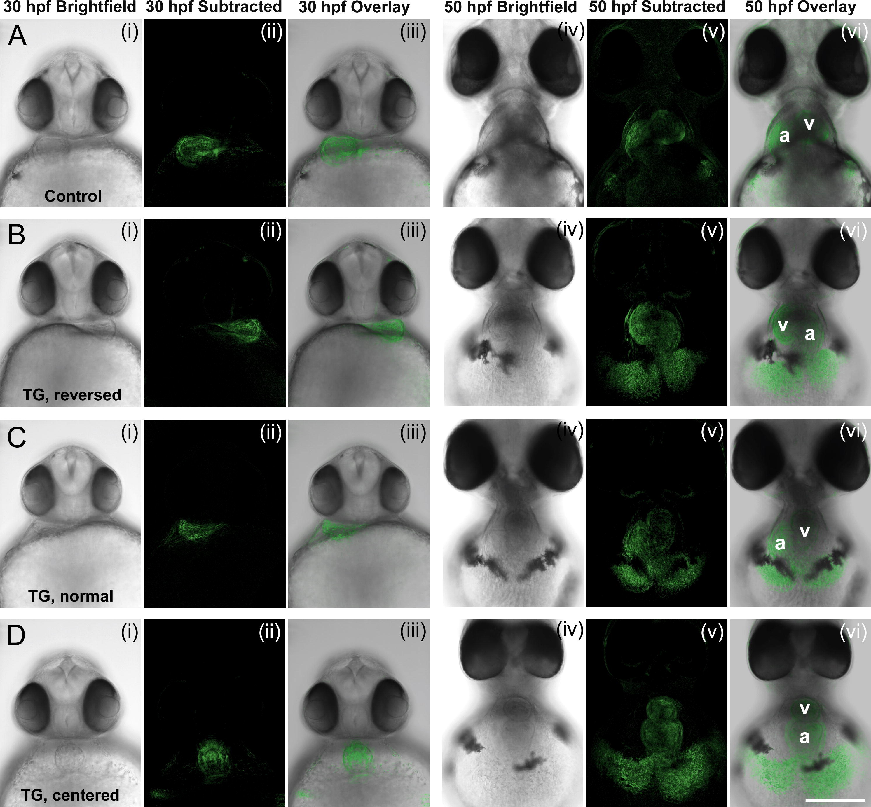
Fig. 2 Suppression of the ER Ca2+ pump during gastrulation affects heart laterality. (A) Control embryos, treated with 1 μl/ml DMSO from 4 to 6 hpf, and imaged at 30 and 50 hpf. The heart is positioned on the left at 30 hpf and shows a D-loop at 50 hpf, with the atrium (a) located on the left and the ventricle (v) on the right. (B–D) Embryos treated with 0.5 μM thapsigargin from 4 to 6 hpf and imaged at 30 and 50 hpf. Thapsigargin-treated embryos display a randomized cardiac orientation, including a reversed orientation or L-loop (B) a normal orientation or D-loop (C), and a centralized orientation or no-loop (D). Each embryo is shown in a bright field image (i, iv), pseudo-colored subtractive image (ii, v), and overlay of the bright field image and subtractive image (iii, vi). The images are shown as collected on an inverted microscope, with embryo’s left on the left side of the image, and the embryo’s right on the right side of the image. Scale bar = 200 μm.
Reprinted from Mechanisms of Development, 125(5-6), Kreiling, J.A., Balantac, Z.L., Crawford, A.R., Ren, Y., Toure, J., Zchut, S., Kochilas, L., and Creton, R., Suppression of the endoplasmic reticulum calcium pump during zebrafish gastrulation affects left-right asymmetry of the heart and brain, 396-410, Copyright (2008) with permission from Elsevier. Full text @ Mech. Dev.