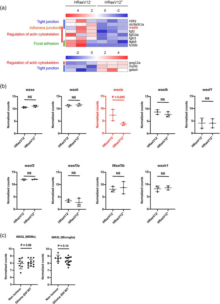
Image
Figure Caption

FIGURE 4

The actin nucleation promoting factor wasla is less expressed in microglia from HRasV12+ larvae. (a) Heatmap of differentially expressed (DE) genes (FDR < 0.05, Fold Change > |2|) from microglia transcriptome comparisons between 5 dpf HRasV12 and HRasV12+ larval brains (11 genes), belonging to zebrafish actin cytoskeleton KEGG pathways [Regulation of actin cytoskeleton, Focal adhesion, Adherens junction, Tight junction and mTOR signaling pathway]. The actin nucleation promoting factor wasla is the top ranked gene of the list. See also Table S3. (b) Dot plots of normalized transformed read counts of the representative WASP family genes. Black plots represent non‐DE genes whereas, red plots correspond to DE genes at 5 dpf between control and HRasV12+ conditions. (b) The means ± SD of three independent experiments are plotted. (c) Dot plots of normalized transformed read counts of WASL gene expression of monocyte‐derived macrophages (MDMs) and microglia in human non tumor and glioma IDH WT brain samples (Klemm et al., 2020)

Figure Data
Acknowledgments
This image is the copyrighted work of the attributed author or publisher, and ZFIN has permission only to display this image to its users. Additional permissions should be obtained from the applicable author or publisher of the image. Full text @ Glia