Fig. 8
Fig. 8
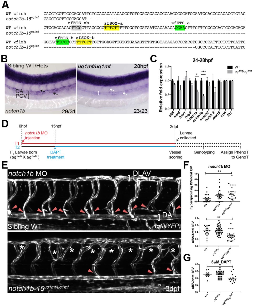
Loss of endogenous notch1b-15 compromises artery formation and reduces the endogenous notch1b transcript level. (A) The deleted region (dashed line) of notch1b-15 mutant allele uq1mf, which includes both the zfSOX-a and zfSOX-b sites (yellow). (B) F2 notch1b-15uq1mf/uq1mf has reduced notch1b expression in dorsal aorta and intersomitic vessels (white arrows) as compared with sibling WT and heterozygotes (black arrows) at 26-28 hpf. The number of embryos showing the illustrated phenotype among the total examined is indicated. (C) Quantitative PCR on FACS-sorted endothelial populations at 24-28 hpf, showing that F3 notch1b-15uq1mf/uq1mf fish have lower notch1b expression than WT fish. Expression is relative to kdrl and flt1. Mean±s.e.m. n=6 (uq1mf/uq1mf) and n=8 (WT) independent sorts, where each sort was pooled from 60-100 larvae;. *P<0.05, ***P<0.001 (t-test). (D) The treatment regime conducted to characterise the vascular phenotype of the uq1mf/+ cross. In treatment 1 (T1, red), notch1b MO was injected at the 1-2 cell stage. The developing vasculature of each embryo was then analysed blindly at 3 dpf. After scoring, genotypes were assigned to each larva. In treatment 2 (T2, blue), larvae from the uq1mf/+ cross were treated with or without DAPT (5 µM) from 15-16 hpf until 3 dpf. Vessels of these treated larvae were blindly scored prior to genotyping, as reported for T1. (E) At 3 dpf, notch1b-15uq1mf/uq1mf notch1b morphants frequently showed ectopic sprouting in between the intersomitic vessels (asterisks) as compared with sibling WT notch1b morphants. Mutants also show loss of arterial connections (red arrowheads) between the dorsal longitudinal anastomotic vessel (DLAV) and dorsal aorta (DA), as indicated by the loss of YFP expression in the tg(flt1:YFP) background. (F) (Top) Quantification of hypersprouting ISV number in individual notch1b morphants labelled by tg(flt1:YFP) at 3 dpf. Mean±s.e.m. Sibling WT (+/+), n=20; heterozygote (uq1mf/+), n=32; homozygous mutant (uq1mf/uq1mf), n=21. (Bottom) Quantification of YFP-positive intersomitic vessels that connect between DLAV and DA in individual notch1b morphants labelled by tg(flt1:YFP) at 3 dpf. Mean±s.e.m. Sibling WT, n=20; heterozygote, n=32; homozygous mutant, n=21. **P<0.005 (Mann–Whitney U-test). (G) Quantification of YFP-positive intersomitic vessels that connect between DLAV and DA in individual embryos treated with 5 μM DAPT at 3 dpf. Vessels are labelled by tg(flt1:YFP). Mean±s.e.m. Sibling WT, n=9; heterozygote, n=25; homozygous mutant, n=14. *P<0.05 (Mann–Whitney U-test).

