
Fig. S4
|
Fig. S4
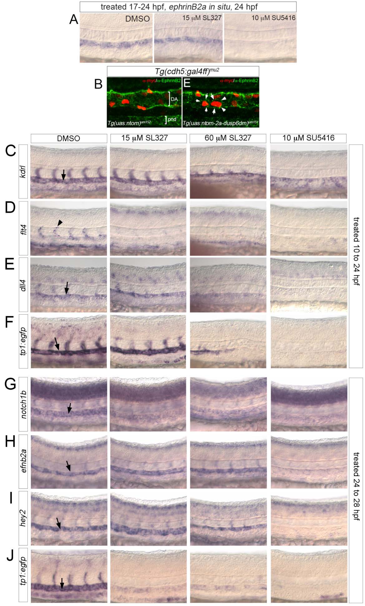
Artery marker gene expression following ERK inhibition. (A) Expresion of efnb2a in embryos treated with indicated compounds from 17 hpf (15 somite stage) to 25 hpf. (B) Confocal images of embryos at 30 hpf bearing indicated transgenes. Myc staining of nTom cells is red and EphrinB2 is in green. Arrows denote dusp6dm-expressing cell expressing EphrinB2. The stained structure ventral to the dorsal aorta is the pronephric duct (pnd) (C-J) Whole mount in situ hybridization with indicated riboprobes. Chemical treatment is indicated at top, stage of treatment at right. Embryos were fixed immediately following treatment. Arrows indicate position of the dorsal aorta.