
Fig. 2
|
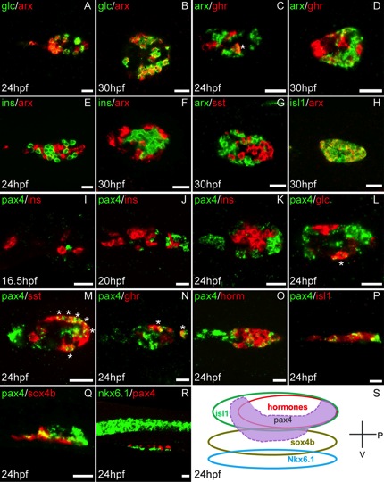
Fig. 2 Identification of pax4 and arx expressing cells by double fluorescent in situ hybridization. All images are confocal optical sections of the dorsal pancreatic bud with anterior part to the left. The probes and the developmental stages are respectively indicated at the top and the bottom in each image. All views are ventral, except images P, Q, R and S which are lateral views. arx transcripts are detected in almost all glucagon+ (A, B), in some ghrelin+ (C, D) cells and in many isl1+ cells (H); absence of co-staining between arx and insulin (E, F) and somatostatin (G). pax4 transcripts are not detected in insulin+ (I - K) and most glucagon+ cells (L). pax4 is expressed in many somatostatin (M) and a few ghrelin (N) expressing cells. Partial co-staining of pax4 probe with a cocktail of insulin, glucagon, somatostatin and ghrelin probes (horm)(O), with isl1 (P) and sox4b (Q) probes. Absence of co-staining between pax4 and nkx6.1 (R). S: Schematic representation of the multi-layer organization of the pancreatic dorsal bud at 24 hpf including pax4 expression. Scale bar = 20 μm. astérisks (*) indicate double positive cells.