
Fig. 6
- ID
- ZDB-IMAGE-100806-104
- Genes
- Publication
- Talbot et al., 2010 - hand2 and Dlx genes specify dorsal, intermediate and ventral domains within zebrafish pharyngeal arches
- All Figures
- Figures for Talbot et al., 2010
|
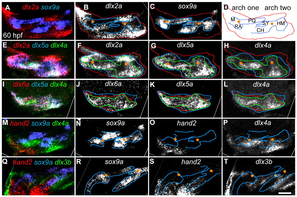
Fig. 6 The patterning domains delineated by Dlx genes and hand2 can be connected to specific pre-skeletal shapes at 60 hpf. (A-P) Lateral views (anterior to the left, dorsal upwards) of RNA in situs confocal sections illustrate differences in dorsal expression boundaries, whereas ventral views (Q-T) (anterior towards the left, lateral upwards) illustrate ventral boundaries. (A-P) Merge of indicated markers is shown in the left column, whereas the other columns show single channels taken from the merge. Joints in the first two arches are indicated by asterisks. Confocal sections in I-L are lateral to cartilages, making the locations of underlying joints difficult to determine. Outlines in single channel panels follow the color schemes shown in the left column. CH, ceratohyal cartilage; HM, hyomandibular region; M, Meckel′s cartilage; PQ, palatoquadrate cartilage; RA, retroarticular process; SY, symplectic region. Scale bar: 50 μm.