|
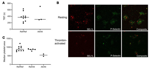
Clinical phenotyping and analysis of platelets from affected horses. (A) TBTs using horses that were Ref/Ref or Alt/Alt for the SEL1L c.1810A>G p.Ile604Val variant found no significant difference between genotypes by 2-tailed Student’s t test. n = 10 Ref/Ref and 3 Alt/Alt. (B) SEL1L (red) localization in relation to P selectin (green) in (top) resting permeabilized platelets and (bottom) thrombin-activated platelet surface of horses that are Ref/Ref for SEL1L c.1810A>G p.Ile604Val. SEL1L did not localize to the surface in resting permeabilized platelets (top) but did localize to the surface in thrombin-activated platelets (bottom). n = 2. Original magnification, ×100. (C) Collagen spreading assay comparing median platelet area to genotype. There was a substantial difference between homozygous Ref and Alt genotypes. n = 11 Ref/Ref, 5 Ref/Alt, and 2 Alt/Alt.
|