|
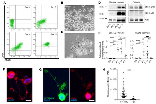
SEL1L is expressed in human cord blood megakaryocytes and platelets. (A) Flow cytometry analyses of human megakaryocytic surface markers from differentiated cord blood. CD61hiCD42bhi megakaryocytes are observed after 2 weeks of differentiation. (B) Phase contrast microscopy of mature human megakaryocytes and (C) proplatelets. Scale bars: 50 μm. (D) Representative Western blot analysis of SEL1L during megakaryopoiesis and in platelets from peripheral blood. (E) Densitometric analyses of SEL1L isoforms (n = 7, results are presented as mean ± SD). **P < 0.01, ***P < 0.001, and ****P < 0.0001 by 1-way ANOVA with post hoc pairwise comparisons. (F and G) Immunofluorescence microscopy of megakaryocytes extending proplatelets. Scale bars: 30 μm. (H) Analysis of SEL1L fluorescence intensity in the megakaryocytic cell body and proplatelet tips. ****P < 0.0001 by 2-tailed Student’s t test.
|