|
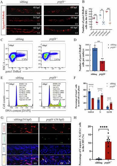
prpf4 deficiency reduces the number of erythrocytes in zebrafish. A, B The number of erythrocytes in the CHT region of siblings and prpf4−/− embryos was assessed using confocal microscopy in Tg(gata1:DsRed) transgenic zebrafish. Scale bar: 100 µm. C, D FACS analysis of GATA1+ erythrocytes from siblings and prpf4−/− mutants at 48 hpf. C In prpf4 mutants, the proportion of GATA1+ erythrocytes was reduced (30 embryos per group). D Quantitative analysis of GATA1+ erythrocyte numbers in individual embryos. E Cell cycle analysis of GATA1+ erythrocytes from siblings and prpf4−/− mutants at 48 hpf (30 embryos per group). F Quantification of the percentage of cells in each cell cycle phase. G, H Confocal microscopy analysis of apoptosis in GATA1+ erythrocytes in the CHT region of siblings and prpf4−/− embryos at 54 hpf. G Immunofluorescence staining showing GATA1 (red), active Caspase-3 (green), and DAPI (blue). Arrows indicate apoptotic GATA1+ cells. Scale bar: 50 µm. H Quantitative analysis shows a significantly higher proportion of apoptotic GATA1+ cells in the CHT region of prpf4−/− embryos compared with siblings.
|