|
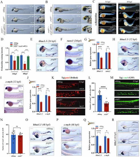
The prpf4 mutation disrupted erythropoiesis in zebrafish. A, B Embryos with prpf4 deficiency exhibit abnormal morphology. A At 36 hpf, prpf4 mutants exhibited a slight tail curvature and a slightly paler coloration in the ventral yolk sac region (a1–a2’). The abnormal morphology of prpf4 mutants was rescued by the injection of prpf4 mRNA (a2–a3’). B At 60 hpf, prpf4 mutants exhibited bulging ventricle, paler coloration in the cardiac region, tail curvature, and a noticeably smaller head (b1–b2’). The injection of prpf4 mRNA effectively rescued the abnormal morphology of prpf4 mutants (b2–b3’). The white arrows indicate the ventral yolk sac region of the embryos. C, D Detection of hemoglobin levels in siblings and prpf4 mutants at 36 hpf and 48 hpf using o-dianisidine staining. E–M Analysis of erythroid and hematopoietic progenitor markers in prpf4−/− embryos. E WISH showing hbae1.1 expression in siblings and prpf4−/− embryos at 26 hpf. F WISH analysis of runx1 expression in siblings and prpf4−/− embryos at 26 hpf. G RT-qPCR analysis of dissected tail regions from 26 hpf embryos. Compared with siblings (set as 1), prpf4−/− embryos showed reduced hbae1.1 expression (0.68-fold), while runx1 expression remained unchanged. H WISH showing hbae1.1 expression in siblings and prpf4−/− embryos at 32 hpf. I WISH showing c-myb expression in siblings and prpf4−/− embryos at 32 hpf. J RT-qPCR analysis of dissected tail regions from 32 hpf embryos. Compared with siblings (set as 1), hbae1.1 expression was reduced in prpf4−/− embryos (0.59-fold), whereas c-myb expression showed no significant difference. K Confocal images of Tg(gata1:DsRed) embryos at 32 hpf showing erythroid cells in the CHT region. L Quantification of Gata1⁺ cells in the CHT revealed a significant reduction in prpf4−/− embryos compared with siblings. M Confocal images of Tg(c-myb:GFP) embryos at 32 hpf showing HSPCs in the AGM region. N Quantification of C-myb⁺ HSPCs in the AGM showed no significant difference between prpf4−/− embryos and siblings. O WISH showing hbae1.1 expression in siblings and prpf4−/− embryos at 48 hpf. P WISH showing c-myb expression in siblings and prpf4−/− embryos at 48 hpf. Q RT-qPCR analysis of dissected tail regions from 48 hpf embryos. prpf4−/− embryos exhibited decreased hbae1.1 expression (0.43-fold compared with siblings set as 1), while c-myb expression was reduced to 0.89-fold relative to siblings. R WISH showing hbbe3 expression patterns in prpf4 mutants and siblings at 48 hpf. Scale bars: 200 μm unless otherwise indicated; 100 μm in (K, M).
|