
Fig. 6
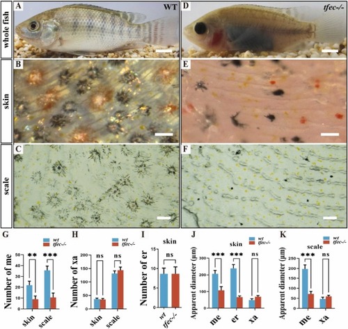
Apparent diameter of melanophores and erythrophores were significantly reduced in tfec−/− mutants at 180 dpf. (A-C) The skin and scales of wild-type fish were covered by dendritic melanophores, dendritic erythrophores, round xanthophores, and shiny iridophores. (D-F) Punctuated melanophores, erythrophores and xanthophores while no iridophores were observed on the skin and scales in tfec−/− mutants. (G) Statistical results of the number of melanophores on the back skin and scale of the fish (n = 6 fish). (H) Statistical results of the number of xanthophores on the back skin and scale of the fish (n = 6 fish). (I) Statistical results of the number of erythrophores on the back skin of the fish (n = 6 fish). (J) Statistical results of the apparent diameter of melanophores, xanthophores and erythrophores on the back skin of the fish (n = 6 fish). (K) Statistical results of the apparent diameter of melanophores, xanthophores and erythrophores on the back scale of the fish (n = 6 fish). Qualification of pigment cells in G-K was performed on the skin (at the same magnification and position for B and E) and scales (at the same magnification and position for C and F) of the fish. Scale bar in A, D, 2 cm; Scale bar in B, C, E, F, 200 μm. |
Reprinted from New biotechnology, 89, Liang, G., Lu, B., Dai, S., Li, M., Yao, J., Liu, H., Liu, X., Liu, X., Wang, D., Creation of colorless transparent tilapia using CRISPR/Cas9 mediated multi-gene mutation, 163-176, Copyright (2025) with permission from Elsevier. Full text @ N. Biotechnol.