
|
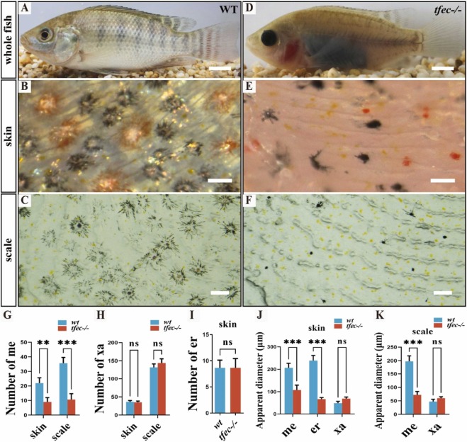
Fig. 6 Apparent diameter of melanophores and erythrophores were significantly reduced in tfec−/− mutants at 180 dpf. (A-C) The skin and scales of wild-type fish were covered by dendritic melanophores, dendritic erythrophores, round xanthophores, and shiny iridophores. (D-F) Punctuated melanophores, erythrophores and xanthophores while no iridophores were observed on the skin and scales in tfec−/− mutants. (G) Statistical results of the number of melanophores on the back skin and scale of the fish (n = 6 fish). (H) Statistical results of the number of xanthophores on the back skin and scale of the fish (n = 6 fish). (I) Statistical results of the number of erythrophores on the back skin of the fish (n = 6 fish). (J) Statistical results of the apparent diameter of melanophores, xanthophores and erythrophores on the back skin of the fish (n = 6 fish). (K) Statistical results of the apparent diameter of melanophores, xanthophores and erythrophores on the back scale of the fish (n = 6 fish). Qualification of pigment cells in G-K was performed on the skin (at the same magnification and position for B and E) and scales (at the same magnification and position for C and F) of the fish. Scale bar in A, D, 2 cm; Scale bar in B, C, E, F, 200 μm.
Reprinted from New biotechnology, 89, Liang, G., Lu, B., Dai, S., Li, M., Yao, J., Liu, H., Liu, X., Liu, X., Wang, D., Creation of colorless transparent tilapia using CRISPR/Cas9 mediated multi-gene mutation, 163-176, Copyright (2025) with permission from Elsevier. Full text @ N. Biotechnol.