
Fig. 6
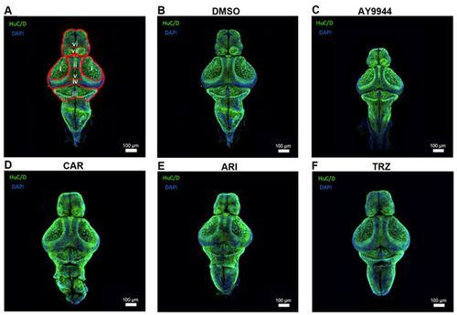
(A–F) Immunohistochemical evaluation of zebrafish brain following exposure to AY9944 and psychopharmaceuticals. Immunofluorescence of HuC/D (green, pan-neuronal stain) and DAPI (blue, nuclear). (A) Measurements of immunolabeled zebrafish brains were taken from the dorsal view. Measurement i: outlines the optic tectum. Measurement ii: measures the distance between tectums and spans from the most medial side of the left optic tectum to the most medial side of the right optic tectum. Measurement iii: outlines the cerebellum. Measurement iv: outlines the midbrain. Measurement v: outlines the mesencephalon between the optic tectums. Measurement vi: measures forebrain length from the most lateral side on the left side of the forebrain to the most lateral side on the right side of the forebrain. Measurement vii: outlines the forebrain. (B) VEH-treated zebrafish brain. (C) AY9944-treated zebrafish brain. (D) CAR-treated zebrafish brain. (E) ARI-treated zebrafish brain. (F) TRZ-treated zebrafish brain. Images were taken at a magnification of 10X and enhanced for viewing. Analyses were performed on raw images. Scale bar: 100 µm. |